6MSF

F6 APTAMER MS2 COAT PROTEIN COMPLEX


Experimental Data Snapshot

  • Method: X-RAY DIFFRACTION
  • Resolution: 2.80 Å
  • R-Value Free: 0.201 
  • R-Value Work: 0.195 
  • R-Value Observed: 0.195 

wwPDB Validation   3D Report Full Report


This is version 1.3 of the entry. See complete history


Literature

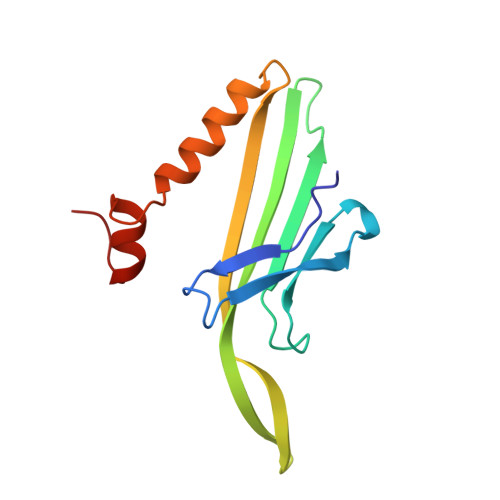
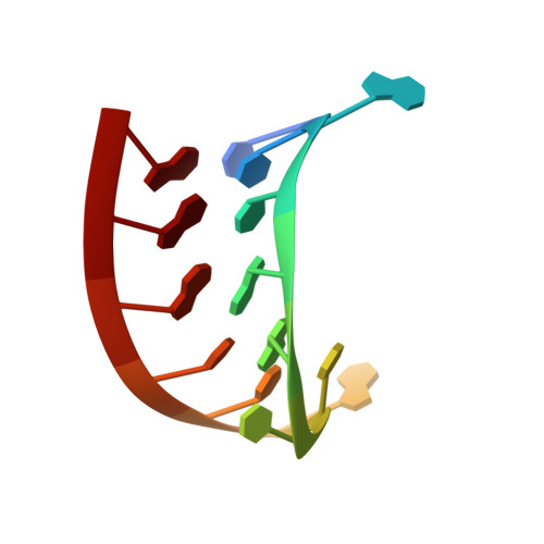
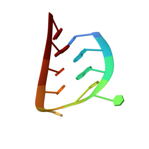
Crystal structure of an RNA aptamer-protein complex at 2.8 A resolution.

Convery, M.A.Rowsell, S.Stonehouse, N.J.Ellington, A.D.Hirao, I.Murray, J.B.Peabody, D.S.Phillips, S.E.Stockley, P.G.

(1998) Nat Struct Biol 5: 133-139

  • DOI: https://doi.org/10.1038/nsb0298-133
  • Primary Citation of Related Structures:  
    6MSF

  • PubMed Abstract: 

    The crystal structure, at 2.8 A resolution, of an RNA aptamer bound to bacteriophage MS2 coat protein has been determined. It provides an opportunity to compare the interactions of MS2 coat protein and wild type operator with those of an aptamer, whose secondary structure differs from the wild type RNA in having a three-base loop (compared to a tetraloop) and an additional base pair between this loop and the sequence-specific recognition element in the stem. The RNA binds in the same location on the coat protein as the wild type operator and maintains many of the same RNA-protein interactions. In order to achieve this, the RNA stem loop undergoes a concerted rearrangement of the 3' side while leaving the 5' side and the loop interactions largely unchanged, illustrating the ability of RNA to present similar molecular recognition surfaces from distinct primary and secondary structures.


  • Organizational Affiliation

    School of Biochemistry and Molecular Biology, North of England Structural Biology Centre, University of Leeds, UK.


Macromolecules

Find similar proteins by:  (by identity cutoff)  |  3D Structure
Entity ID: 3
MoleculeChains Sequence LengthOrganismDetailsImage
PROTEIN (MS2 PROTEIN CAPSID)C [auth A],
D [auth B],
E [auth C]
129Escherichia phage MS2Mutation(s): 0 
Gene Names: COAT PROTEIN
UniProt
Find proteins for P03612 (Escherichia phage MS2)
Explore P03612 
Go to UniProtKB:  P03612
Entity Groups  
Sequence Clusters30% Identity50% Identity70% Identity90% Identity95% Identity100% Identity
UniProt GroupP03612
Sequence Annotations
Expand
  • Reference Sequence

Find similar nucleic acids by:  Sequence   |   3D Structure  

Entity ID: 1
MoleculeChains LengthOrganismImage
RNA (5'-R(*CP*CP*AP*CP*AP*GP*UP*CP*AP*CP*UP*GP*GP*G)-3')A [auth R]14N/A
Sequence Annotations
Expand
  • Reference Sequence

Find similar nucleic acids by:  Sequence   |   3D Structure  

Entity ID: 2
MoleculeChains LengthOrganismImage
RNA (5'-R(*CP*AP*GP*UP*CP*AP*CP*UP*GP*G)-3')B [auth S]10N/A
Sequence Annotations
Expand
  • Reference Sequence
Experimental Data & Validation

Experimental Data

  • Method: X-RAY DIFFRACTION
  • Resolution: 2.80 Å
  • R-Value Free: 0.201 
  • R-Value Work: 0.195 
  • R-Value Observed: 0.195 
  • Space Group: H 3 2
Unit Cell:
Length ( Å )Angle ( ˚ )
a = 287.58α = 90
b = 287.58β = 90
c = 652.19γ = 120
Software Package:
Software NamePurpose
CCP4model building
X-PLORrefinement
MOSFLMdata reduction
CCP4data scaling
CCP4phasing

Structure Validation

View Full Validation Report


Entry History 

Deposition Data

Revision History  (Full details and data files)

  • Version 1.0: 1998-07-08
    Type: Initial release
  • Version 1.1: 2008-05-22
    Changes: Version format compliance
  • Version 1.2: 2011-07-13
    Changes: Version format compliance
  • Version 1.3: 2023-08-02
    Changes: Database references, Derived calculations, Refinement description