6GPB

REFINED CRYSTAL STRUCTURE OF THE PHOSPHORYLASE-HEPTULOSE 2-PHOSPHATE-OLIGOSACCHARIDE-AMP COMPLEX


Experimental Data Snapshot

  • Method: X-RAY DIFFRACTION
  • Resolution: 2.86 Å
  • R-Value Work: 0.201 

wwPDB Validation   3D Report Full Report


This is version 2.0 of the entry. See complete history


Literature

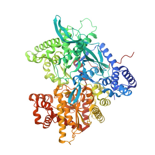
Refined crystal structure of the phosphorylase-heptulose 2-phosphate-oligosaccharide-AMP complex.

Johnson, L.N.Acharya, K.R.Jordan, M.D.McLaughlin, P.J.

(1990) J Mol Biol 211: 645-661

  • DOI: https://doi.org/10.1016/0022-2836(90)90271-M
  • Primary Citation of Related Structures:  
    6GPB

  • PubMed Abstract: 

    The crystal structure of phosphorylase b-heptulose 2-phosphate complex with oligosaccharide and AMP bound has been refined by molecular dynamics and crystallographic least-squares with the program XPLOR. Shifts in atomic positions of up to 4 A from the native enzyme structure were correctly determined by the program without manual intervention. The final crystallographic R value for data between 8 and 2.86 A resolution is 0.201, and the overall root-mean-square difference between the native and complexed structure is 0.58 A for all protein atoms. The results confirm the previous observation that there is a direct hydrogen bond between the phosphate of heptulose 2-phosphate and the pyridoxal phosphate 5'-phosphate group. The close proximity of the two phosphates is stabilized by an arginine residue, Arg569, which shifts from a site buried in the protein to a position where it can make contact with the product phosphate. There is a mutual interchange in position between the arginine and an acidic group, Asp283. These movements represent the first stage of the allosteric response which converts the catalytic site from a low to a high-affinity binding site. Communication of these changes to other sites is prevented in the crystal by the lattice forces, which also form the subunit interface. The constellation of groups in the phosphorylase transition state analogue complex provides a structural basis for understanding the catalytic mechanism in which the cofactor pyridoxal phosphate 5'-phosphate group functions as a general acid to promote attack by the substrate phosphate on the glycosidic bond when the reaction proceeds in the direction of glycogen degradation. In the direction of glycogen synthesis, stereoelectronic effects contribute to the cleavage of the C-1-O-1 bond. In both reactions the substrate phosphate plays a key role in transition state stabilization. The details of the oligosaccharide, maltoheptaose, interactions with the enzyme at the glycogen storage site are also described.


  • Organizational Affiliation

    Laboratory of Molecular Biophysics, Oxford, U.K.


Macromolecules
Find similar proteins by:  (by identity cutoff)  |  3D Structure
Entity ID: 1
MoleculeChains Sequence LengthOrganismDetailsImage
GLYCOGEN PHOSPHORYLASE B842Oryctolagus cuniculusMutation(s): 0 
EC: 2.4.1.1
UniProt
Find proteins for P00489 (Oryctolagus cuniculus)
Explore P00489 
Go to UniProtKB:  P00489
Entity Groups  
Sequence Clusters30% Identity50% Identity70% Identity90% Identity95% Identity100% Identity
UniProt GroupP00489
Sequence Annotations
Expand
  • Reference Sequence
Oligosaccharides

Help

Entity ID: 2
MoleculeChains Length2D Diagram Glycosylation3D Interactions
alpha-D-glucopyranose-(1-4)-alpha-D-glucopyranose-(1-4)-alpha-D-glucopyranose-(1-4)-alpha-D-glucopyranose-(1-4)-alpha-D-glucopyranose
B
5N/A
Glycosylation Resources
GlyTouCan:  G50146AM
GlyCosmos:  G50146AM
Small Molecules
Ligands 3 Unique
IDChains Name / Formula / InChI Key2D Diagram3D Interactions
AMP
Query on AMP

Download Ideal Coordinates CCD File 
E [auth A],
F [auth A]
ADENOSINE MONOPHOSPHATE
C10 H14 N5 O7 P
UDMBCSSLTHHNCD-KQYNXXCUSA-N
H2P
Query on H2P

Download Ideal Coordinates CCD File 
C [auth A]1-deoxy-2-O-phosphono-alpha-D-gluco-hept-2-ulopyranose
C7 H15 O9 P
QZBAZODTRUGOQS-XUUWZHRGSA-N
PLP
Query on PLP

Download Ideal Coordinates CCD File 
D [auth A]PYRIDOXAL-5'-PHOSPHATE
C8 H10 N O6 P
NGVDGCNFYWLIFO-UHFFFAOYSA-N
Binding Affinity Annotations 
IDSourceBinding Affinity
H2P PDBBind:  6GPB Ki: 1.40e+4 (nM) from 1 assay(s)
Biologically Interesting Molecules (External Reference) 1 Unique
Experimental Data & Validation

Experimental Data

  • Method: X-RAY DIFFRACTION
  • Resolution: 2.86 Å
  • R-Value Work: 0.201 
  • Space Group: P 43 21 2
Unit Cell:
Length ( Å )Angle ( ˚ )
a = 128.5α = 90
b = 128.5β = 90
c = 116.3γ = 90
Software Package:
Software NamePurpose
X-PLORrefinement

Structure Validation

View Full Validation Report


Entry History 

Deposition Data

Revision History  (Full details and data files)

  • Version 1.0: 1992-10-15
    Type: Initial release
  • Version 1.1: 2008-03-27
    Changes: Version format compliance
  • Version 1.2: 2011-07-13
    Changes: Derived calculations, Version format compliance
  • Version 1.3: 2017-11-29
    Changes: Derived calculations, Other
  • Version 2.0: 2020-07-29
    Type: Remediation
    Reason: Carbohydrate remediation
    Changes: Advisory, Atomic model, Data collection, Database references, Derived calculations, Structure summary