4H82

Crystal structure of mutant MMP-9 catalytic domain in complex with a twin inhibitor.


Experimental Data Snapshot

  • Method: X-RAY DIFFRACTION
  • Resolution: 1.90 Å
  • R-Value Free: 0.241 
  • R-Value Work: 0.207 
  • R-Value Observed: 0.209 

wwPDB Validation   3D Report Full Report


Ligand Structure Quality Assessment 


This is version 1.5 of the entry. See complete history


Literature

Crystallization of bi-functional ligand protein complexes.

Antoni, C.Vera, L.Devel, L.Catalani, M.P.Czarny, B.Cassar-Lajeunesse, E.Nuti, E.Rossello, A.Dive, V.Stura, E.A.

(2013) J Struct Biol 182: 246-254

  • DOI: https://doi.org/10.1016/j.jsb.2013.03.015
  • Primary Citation of Related Structures:  
    4H1Q, 4H2E, 4H30, 4H3X, 4H49, 4H76, 4H82, 4H84, 4HMA, 4I03

  • PubMed Abstract: 

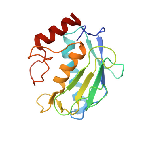
    Homodimerization is important in signal transduction and can play a crucial role in many other biological systems. To obtaining structural information for the design of molecules able to control the signalization pathways, the proteins involved will have to be crystallized in complex with ligands that induce dimerization. Bi-functional drugs have been generated by linking two ligands together chemically and the relative crystallizability of complexes with mono-functional and bi-functional ligands has been evaluated. There are problems associated with crystallization with such ligands, but overall, the advantages appear to be greater than the drawbacks. The study involves two matrix metalloproteinases, MMP-12 and MMP-9. Using flexible and rigid linkers we show that it is possible to control the crystal packing and that by changing the ligand-enzyme stoichiometric ratio, one can toggle between having one bi-functional ligand binding to two enzymes and having the same ligand bound to each enzyme. The nature of linker and its point of attachment on the ligand can be varied to aid crystallization, and such variations can also provide valuable structural information about the interactions made by the linker with the protein. We report here the crystallization and structure determination of seven ligand-dimerized complexes. These results suggest that the use of bi-functional drugs can be extended beyond the realm of protein dimerization to include all drug design projects.


  • Organizational Affiliation

    CEA, iBiTec-S, Service d'Ingénierie Moléculaire des Protéines-SIMOPRO, Gif-sur-Yvette F-91191, France. antoni.claudia@cea.fr


Macromolecules
Find similar proteins by:  (by identity cutoff)  |  3D Structure
Entity ID: 1
MoleculeChains Sequence LengthOrganismDetailsImage
Matrix metalloproteinase-9
A, B, C, D
160Homo sapiensMutation(s): 1 
Gene Names: CLG4BMMP9
EC: 3.4.24.35
UniProt & NIH Common Fund Data Resources
Find proteins for P14780 (Homo sapiens)
Explore P14780 
Go to UniProtKB:  P14780
PHAROS:  P14780
GTEx:  ENSG00000100985 
Entity Groups  
Sequence Clusters30% Identity50% Identity70% Identity90% Identity95% Identity100% Identity
UniProt GroupP14780
Sequence Annotations
Expand
  • Reference Sequence
Small Molecules
Ligands 7 Unique
IDChains Name / Formula / InChI Key2D Diagram3D Interactions
L29
Query on L29

Download Ideal Coordinates CCD File 
EA [auth D],
J [auth A]
N,N'-bis(2-{(biphenyl-4-ylsulfonyl)[(2R)-1-(hydroxyamino)-3-methyl-1-oxobutan-2-yl]amino}ethyl)benzene-1,3-dicarboxamide (non-preferred name)
C46 H52 N6 O10 S2
JPMXJRBHCOULJI-NCRNUEESSA-N
PEG
Query on PEG

Download Ideal Coordinates CCD File 
BA [auth C]
KA [auth D]
U [auth C]
V [auth C]
X [auth C]
BA [auth C],
KA [auth D],
U [auth C],
V [auth C],
X [auth C],
Y [auth C],
Z [auth C]
DI(HYDROXYETHYL)ETHER
C4 H10 O3
MTHSVFCYNBDYFN-UHFFFAOYSA-N
GOL
Query on GOL

Download Ideal Coordinates CCD File 
AA [auth C]GLYCEROL
C3 H8 O3
PEDCQBHIVMGVHV-UHFFFAOYSA-N
PGO
Query on PGO

Download Ideal Coordinates CCD File 
CA [auth D],
JA [auth D],
LA [auth D]
S-1,2-PROPANEDIOL
C3 H8 O2
DNIAPMSPPWPWGF-VKHMYHEASA-N
PPI
Query on PPI

Download Ideal Coordinates CCD File 
W [auth C]PROPANOIC ACID
C3 H6 O2
XBDQKXXYIPTUBI-UHFFFAOYSA-N
ZN
Query on ZN

Download Ideal Coordinates CCD File 
DA [auth D]
E [auth A]
F [auth A]
FA [auth D]
K [auth B]
DA [auth D],
E [auth A],
F [auth A],
FA [auth D],
K [auth B],
L [auth B],
P [auth C],
Q [auth C]
ZINC ION
Zn
PTFCDOFLOPIGGS-UHFFFAOYSA-N
CA
Query on CA

Download Ideal Coordinates CCD File 
G [auth A]
GA [auth D]
H [auth A]
HA [auth D]
I [auth A]
G [auth A],
GA [auth D],
H [auth A],
HA [auth D],
I [auth A],
IA [auth D],
M [auth B],
N [auth B],
O [auth B],
R [auth C],
S [auth C],
T [auth C]
CALCIUM ION
Ca
BHPQYMZQTOCNFJ-UHFFFAOYSA-N
Binding Affinity Annotations 
IDSourceBinding Affinity
L29 BindingDB:  4H82 IC50: 5.5 (nM) from 1 assay(s)
Experimental Data & Validation

Experimental Data

  • Method: X-RAY DIFFRACTION
  • Resolution: 1.90 Å
  • R-Value Free: 0.241 
  • R-Value Work: 0.207 
  • R-Value Observed: 0.209 
  • Space Group: P 1
Unit Cell:
Length ( Å )Angle ( ˚ )
a = 39.9α = 90.03
b = 98.86β = 111.95
c = 47.13γ = 89.98
Software Package:
Software NamePurpose
DNAdata collection
MOLREPphasing
PHENIXrefinement
XDSdata reduction
XDSdata scaling

Structure Validation

View Full Validation Report


Ligand Structure Quality Assessment 


Entry History 

Deposition Data

Revision History  (Full details and data files)

  • Version 1.0: 2013-05-01
    Type: Initial release
  • Version 1.1: 2014-10-08
    Changes: Database references
  • Version 1.2: 2014-10-15
    Changes: Derived calculations
  • Version 1.3: 2015-08-12
    Changes: Database references
  • Version 1.4: 2017-08-09
    Changes: Refinement description, Source and taxonomy
  • Version 1.5: 2023-09-20
    Changes: Data collection, Database references, Derived calculations, Refinement description