4FE5

Crystal structure of the xpt-pbuX guanine riboswitch aptamer domain in complex with hypoxanthine


Experimental Data Snapshot

  • Method: X-RAY DIFFRACTION
  • Resolution: 1.32 Å
  • R-Value Free: 0.229 
  • R-Value Work: 0.196 
  • R-Value Observed: 0.196 

wwPDB Validation   3D Report Full Report


This is version 1.1 of the entry. See complete history


Literature

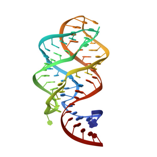
Structure of a natural guanine-responsive riboswitch complexed with the metabolite hypoxanthine.

Batey, R.T.Gilbert, S.D.Montange, R.K.

(2004) Nature 432: 411-415

  • DOI: https://doi.org/10.1038/nature03037
  • Primary Citation of Related Structures:  
    4FE5

  • PubMed Abstract: 

    Riboswitches are genetic regulatory elements found in the 5' untranslated region of messenger RNA that act in the absence of protein cofactors. They are broadly distributed across bacteria and account for the regulation of more than 2% of all genes in Bacillus subtilis, underscoring their importance in the control of cellular metabolism. The 5' untranslated region of many mRNAs of genes involved in purine metabolism and transport contain a guanine-responsive riboswitch that directly binds guanine, hypoxanthine or xanthine to terminate transcription. Here we report the crystal structure at 1.95 A resolution of the purine-binding domain of the guanine riboswitch from the xpt-pbuX operon of B. subtilis bound to hypoxanthine, a prevalent metabolite in the bacterial purine salvage pathway. This structure reveals a complex RNA fold involving several phylogenetically conserved nucleotides that create a binding pocket that almost completely envelops the ligand. Hypoxanthine functions to stabilize this structure and to promote the formation of a downstream transcriptional terminator element, thereby providing a mechanism for directly repressing gene expression in response to an increase in intracellular concentrations of metabolite.


  • Organizational Affiliation

    Department of Chemistry and Biochemistry, 215 UCB, University of Colorado, Boulder, Colorado 80309, USA. robert.batey@colorado.edu


Macromolecules
Find similar nucleic acids by:  (by identity cutoff)  |  3D Structure
Entity ID: 1
MoleculeChains LengthOrganismImage
xpt-pbuX guanine riboswitch aptamer domainA [auth B]67N/A
Sequence Annotations
Expand
  • Reference Sequence
Small Molecules
Ligands 3 Unique
IDChains Name / Formula / InChI Key2D Diagram3D Interactions
NCO
Query on NCO

Download Ideal Coordinates CCD File 
E [auth B]
F [auth B]
G [auth B]
H [auth B]
I [auth B]
E [auth B],
F [auth B],
G [auth B],
H [auth B],
I [auth B],
J [auth B]
COBALT HEXAMMINE(III)
Co H18 N6
DYLMFCCYOUSRTK-UHFFFAOYSA-N
HPA
Query on HPA

Download Ideal Coordinates CCD File 
B
HYPOXANTHINE
C5 H4 N4 O
FDGQSTZJBFJUBT-UHFFFAOYSA-N
ACT
Query on ACT

Download Ideal Coordinates CCD File 
C [auth B],
D [auth B]
ACETATE ION
C2 H3 O2
QTBSBXVTEAMEQO-UHFFFAOYSA-M
Binding Affinity Annotations 
IDSourceBinding Affinity
HPA PDBBind:  4FE5 Kd: 732 (nM) from 1 assay(s)
Experimental Data & Validation

Experimental Data

  • Method: X-RAY DIFFRACTION
  • Resolution: 1.32 Å
  • R-Value Free: 0.229 
  • R-Value Work: 0.196 
  • R-Value Observed: 0.196 
  • Space Group: C 1 2 1
Unit Cell:
Length ( Å )Angle ( ˚ )
a = 133.247α = 90
b = 35.25β = 90.75
c = 41.833γ = 90
Software Package:
Software NamePurpose
HKL-2000data collection
CNSrefinement
HKL-2000data reduction
HKL-2000data scaling
CNSphasing

Structure Validation

View Full Validation Report


Entry History 

Deposition Data

Revision History  (Full details and data files)

  • Version 1.0: 2012-06-27
    Type: Initial release
  • Version 1.1: 2024-02-28
    Changes: Data collection, Database references, Derived calculations