


4DW7
Crystal structure of an active-site mutant of the glycoprotein Erns from the pestivirus BVDV-1 in complex with a CpU dinucleotide
- PDB DOI: https://doi.org/10.2210/pdb4DW7/pdb
- Classification: VIRAL PROTEIN
- Organism(s): Bovine viral diarrhea virus 1-CP7
- Expression System: Drosophila melanogaster
- Mutation(s): Yes
- Deposited: 2012-02-24 Released: 2012-05-23
Experimental Data Snapshot
- Method: X-RAY DIFFRACTION
- Resolution: 3.08 Å
- R-Value Free: 0.245
- R-Value Work: 0.197
- R-Value Observed: 0.200

This is version 2.1 of the entry. See complete history.

Macromolecules
Find similar proteins by:
(by identity cutoff) | 3D Structure
Entity ID: 1 | |||||
|---|---|---|---|---|---|
| Molecule | Chains | Sequence Length | Organism | Details | Image |
| E(rns) glycoprotein | 167 | Bovine viral diarrhea virus 1-CP7 | Mutation(s): 1 Gene Names: Erns | 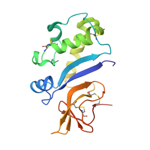 | |
UniProt | |||||
Find proteins for Q96662 (Bovine viral diarrhea virus (strain CP7)) Explore Q96662 Go to UniProtKB: Q96662 | |||||
Entity Groups | |||||
| Sequence Clusters | 30% Identity50% Identity70% Identity90% Identity95% Identity100% Identity | ||||
| UniProt Group | Q96662 | ||||
Sequence AnnotationsExpand | |||||
| |||||
Oligosaccharides
Entity ID: 2 | |||||
|---|---|---|---|---|---|
| Molecule | Chains | Length | 2D Diagram | Glycosylation | 3D Interactions |
| beta-D-mannopyranose-(1-6)-beta-D-mannopyranose-(1-3)-beta-D-mannopyranose-(1-4)-2-acetamido-2-deoxy-beta-D-glucopyranose-(1-4)-2-acetamido-2-deoxy-beta-D-glucopyranose | C | 5 |  | N-Glycosylation | |
Glycosylation Resources | |||||
GlyTouCan: G68945PX GlyCosmos: G68945PX GlyGen: G68945PX | |||||
Entity ID: 3 | |||||
|---|---|---|---|---|---|
| Molecule | Chains | Length | 2D Diagram | Glycosylation | 3D Interactions |
| 2-acetamido-2-deoxy-beta-D-glucopyranose-(1-4)-2-acetamido-2-deoxy-beta-D-glucopyranose | D, F, G | 2 |  | N-Glycosylation | |
Glycosylation Resources | |||||
GlyTouCan: G42666HT GlyCosmos: G42666HT GlyGen: G42666HT | |||||
Entity ID: 4 | |||||
|---|---|---|---|---|---|
| Molecule | Chains | Length | 2D Diagram | Glycosylation | 3D Interactions |
| beta-D-mannopyranose-(1-6)-beta-D-mannopyranose-(1-3)-[alpha-D-mannopyranose-(1-4)-alpha-D-mannopyranose-(1-6)]beta-D-mannopyranose-(1-4)-2-acetamido-2-deoxy-beta-D-glucopyranose-(1-4)-2-acetamido-2-deoxy-beta-D-glucopyranose | E | 7 |  | N-Glycosylation | |
Glycosylation Resources | |||||
GlyTouCan: G21477HL GlyCosmos: G21477HL GlyGen: G21477HL | |||||
Small Molecules
| Ligands 4 Unique | |||||
|---|---|---|---|---|---|
| ID | Chains | Name / Formula / InChI Key | 2D Diagram | 3D Interactions | |
| U Query on U | J [auth A], O [auth B] | URIDINE-5'-MONOPHOSPHATE C9 H13 N2 O9 P DJJCXFVJDGTHFX-XVFCMESISA-N |  | ||
| C Query on C | K [auth A], P [auth B] | CYTIDINE-5'-MONOPHOSPHATE C9 H14 N3 O8 P IERHLVCPSMICTF-XVFCMESISA-N |  | ||
| NAG Query on NAG | H [auth A], I [auth A] | 2-acetamido-2-deoxy-beta-D-glucopyranose C8 H15 N O6 OVRNDRQMDRJTHS-FMDGEEDCSA-N |  | ||
| SO4 Query on SO4 | L [auth A], M [auth A], N [auth A], Q [auth B], R [auth B] | SULFATE ION O4 S QAOWNCQODCNURD-UHFFFAOYSA-L |  | ||
Experimental Data & Validation
Experimental Data
- Method: X-RAY DIFFRACTION
- Resolution: 3.08 Å
- R-Value Free: 0.245
- R-Value Work: 0.197
- R-Value Observed: 0.200
- Space Group: P 65 2 2
Unit Cell:
| Length ( Å ) | Angle ( ˚ ) |
|---|---|
| a = 105.896 | α = 90 |
| b = 105.896 | β = 90 |
| c = 211.794 | γ = 120 |
| Software Name | Purpose |
|---|---|
| SCALA | data scaling |
| PHASER | phasing |
| BUSTER-TNT | refinement |
| PDB_EXTRACT | data extraction |
| XDS | data scaling |
| XDS | data reduction |
| BUSTER | refinement |
Entry History
Deposition Data
- Released Date: 2012-05-23 Deposition Author(s): Krey, T., Bontems, F., Vonrhein, C., Vaney, M.-C., Bricogne, G., Ruemenapf, T., Rey, F.A.
Revision History (Full details and data files)
- Version 1.0: 2012-05-23
Type: Initial release - Version 2.0: 2020-07-29
Type: Remediation
Reason: Carbohydrate remediation
Changes: Advisory, Atomic model, Data collection, Database references, Derived calculations, Structure summary - Version 2.1: 2023-09-13
Changes: Data collection, Database references, Refinement description, Structure summary
