4DVK

Crystal structure of the glycoprotein Erns from the pestivirus BVDV-1 strain NCP-7


Experimental Data Snapshot

  • Method: X-RAY DIFFRACTION
  • Resolution: 2.21 Å
  • R-Value Free: 0.225 
  • R-Value Work: 0.209 
  • R-Value Observed: 0.210 

wwPDB Validation   3D Report Full Report


Ligand Structure Quality Assessment 


This is version 2.0 of the entry. See complete history


Literature

Crystal Structure of the Pestivirus Envelope Glycoprotein E(rns) and Mechanistic Analysis of Its Ribonuclease Activity.

Krey, T.Bontems, F.Vonrhein, C.Vaney, M.C.Bricogne, G.Rumenapf, T.Rey, F.A.

(2012) Structure 20: 862-873

  • DOI: https://doi.org/10.1016/j.str.2012.03.018
  • Primary Citation of Related Structures:  
    4DVK, 4DVL, 4DVN, 4DW3, 4DW4, 4DW5, 4DW7, 4DWA, 4DWC

  • PubMed Abstract: 

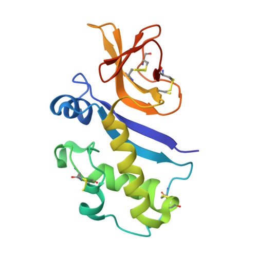
    Pestiviruses, which belong to the Flaviviridae family of RNA viruses, are important agents of veterinary diseases causing substantial economical losses in animal farming worldwide. Pestivirus particles display three envelope glycoproteins at their surface: E(rns), E1, and E2. We report here the crystal structure of the catalytic domain of E(rns), the ribonucleolytic activity of which is believed to counteract the innate immunity of the host. The structure reveals a three-dimensional fold corresponding to T2 ribonucleases from plants and fungi. Cocrystallization experiments with mono- and oligonucleotides revealed the structural basis for substrate recognition at two binding sites previously identified for T2 RNases. A detailed analysis of poly-U cleavage products using (31)P-NMR and size exclusion chromatography, together with molecular docking studies, provides a comprehensive mechanistic picture of E(rns) activity on its substrates and reveals the presence of at least one additional nucleotide binding site.


  • Organizational Affiliation

    Unité de Virologie Structurale, Institut Pasteur, 75015 Paris, France. tkrey@pasteur.fr


Macromolecules
Find similar proteins by:  (by identity cutoff)  |  3D Structure
Entity ID: 1
MoleculeChains Sequence LengthOrganismDetailsImage
E(rns) glycoprotein
A, B
165Bovine viral diarrhea virus 1-CP7Mutation(s): 0 
Gene Names: Erns
UniProt
Find proteins for Q96662 (Bovine viral diarrhea virus (strain CP7))
Explore Q96662 
Go to UniProtKB:  Q96662
Entity Groups  
Sequence Clusters30% Identity50% Identity70% Identity90% Identity95% Identity100% Identity
UniProt GroupQ96662
Sequence Annotations
Expand
  • Reference Sequence
Oligosaccharides

Help

Entity ID: 2
MoleculeChains Length2D Diagram Glycosylation3D Interactions
alpha-D-mannopyranose-(1-4)-alpha-D-mannopyranose-(1-6)-[beta-D-mannopyranose-(1-3)]beta-D-mannopyranose-(1-4)-2-acetamido-2-deoxy-beta-D-glucopyranose-(1-4)-2-acetamido-2-deoxy-beta-D-glucopyranose
C
6N-Glycosylation
Glycosylation Resources
GlyTouCan:  G51778NN
GlyCosmos:  G51778NN
GlyGen:  G51778NN
Entity ID: 3
MoleculeChains Length2D Diagram Glycosylation3D Interactions
2-acetamido-2-deoxy-beta-D-glucopyranose-(1-4)-2-acetamido-2-deoxy-beta-D-glucopyranose
D, F, G
2N-Glycosylation
Glycosylation Resources
GlyTouCan:  G42666HT
GlyCosmos:  G42666HT
GlyGen:  G42666HT
Entity ID: 4
MoleculeChains Length2D Diagram Glycosylation3D Interactions
beta-D-mannopyranose-(1-6)-beta-D-mannopyranose-(1-3)-[alpha-D-mannopyranose-(1-4)-alpha-D-mannopyranose-(1-6)]beta-D-mannopyranose-(1-4)-2-acetamido-2-deoxy-beta-D-glucopyranose-(1-4)-2-acetamido-2-deoxy-beta-D-glucopyranose
E
7N-Glycosylation
Glycosylation Resources
GlyTouCan:  G21477HL
GlyCosmos:  G21477HL
GlyGen:  G21477HL
Small Molecules
Ligands 2 Unique
IDChains Name / Formula / InChI Key2D Diagram3D Interactions
NAG
Query on NAG

Download Ideal Coordinates CCD File 
H [auth A]
I [auth A]
J [auth A]
N [auth B]
O [auth B]
H [auth A],
I [auth A],
J [auth A],
N [auth B],
O [auth B],
P [auth B]
2-acetamido-2-deoxy-beta-D-glucopyranose
C8 H15 N O6
OVRNDRQMDRJTHS-FMDGEEDCSA-N
SO4
Query on SO4

Download Ideal Coordinates CCD File 
K [auth A]
L [auth A]
M [auth A]
Q [auth B]
R [auth B]
K [auth A],
L [auth A],
M [auth A],
Q [auth B],
R [auth B],
S [auth B],
T [auth B],
U [auth B],
V [auth B],
W [auth B]
SULFATE ION
O4 S
QAOWNCQODCNURD-UHFFFAOYSA-L
Experimental Data & Validation

Experimental Data

  • Method: X-RAY DIFFRACTION
  • Resolution: 2.21 Å
  • R-Value Free: 0.225 
  • R-Value Work: 0.209 
  • R-Value Observed: 0.210 
  • Space Group: P 65 2 2
Unit Cell:
Length ( Å )Angle ( ˚ )
a = 106.04α = 90
b = 106.04β = 90
c = 211.85γ = 120
Software Package:
Software NamePurpose
SCALAdata scaling
SHARPphasing
SOLOMONphasing
BUSTER-TNTrefinement
PDB_EXTRACTdata extraction
XDSdata scaling
XDSdata reduction
BUSTERrefinement

Structure Validation

View Full Validation Report


Ligand Structure Quality Assessment 


Entry History 

Deposition Data

Revision History  (Full details and data files)

  • Version 1.0: 2012-05-23
    Type: Initial release
  • Version 2.0: 2020-07-29
    Type: Remediation
    Reason: Carbohydrate remediation
    Changes: Advisory, Atomic model, Data collection, Derived calculations, Structure summary