4APX

CRYSTAL STRUCTURE OF MOUSE CADHERIN-23 EC1-2 AND PROTOCADHERIN-15 EC1- 2 FORM I


Experimental Data Snapshot

  • Method: X-RAY DIFFRACTION
  • Resolution: 1.65 Å
  • R-Value Free: 0.193 
  • R-Value Work: 0.161 
  • R-Value Observed: 0.163 

wwPDB Validation   3D Report Full Report


This is version 1.3 of the entry. See complete history


Literature

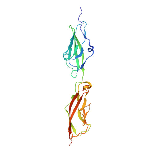
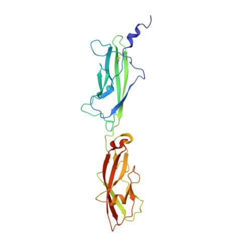
Structure of a Force-Conveying Cadherin Bond Essential for Inner-Ear Mechanotransduction

Sotomayor, M.Weihofen, W.Gaudet, R.Corey, D.P.

(2012) Nature 492: 128

  • DOI: https://doi.org/10.1038/nature11590
  • Primary Citation of Related Structures:  
    4APX, 4AQ8, 4AQA, 4AQE, 4AXW

  • PubMed Abstract: 

    Hearing and balance use hair cells in the inner ear to transform mechanical stimuli into electrical signals. Mechanical force from sound waves or head movements is conveyed to hair-cell transduction channels by tip links, fine filaments formed by two atypical cadherins known as protocadherin 15 and cadherin 23 (refs 4, 5). These two proteins are involved in inherited deafness and feature long extracellular domains that interact tip-to-tip in a Ca(2+)-dependent manner. However, the molecular architecture of this complex is unknown. Here we combine crystallography, molecular dynamics simulations and binding experiments to characterize the protocadherin 15-cadherin 23 bond. We find a unique cadherin interaction mechanism, in which the two most amino-terminal cadherin repeats (extracellular cadherin repeats 1 and 2) of each protein interact to form an overlapped, antiparallel heterodimer. Simulations predict that this tip-link bond is mechanically strong enough to resist forces in hair cells. In addition, the complex is shown to become unstable in response to Ca(2+) removal owing to increased flexure of Ca(2+)-free cadherin repeats. Finally, we use structures and biochemical measurements to study the molecular mechanisms by which deafness mutations disrupt tip-link function. Overall, our results shed light on the molecular mechanics of hair-cell sensory transduction and on new interaction mechanisms for cadherins, a large protein family implicated in tissue and organ morphogenesis, neural connectivity and cancer.


  • Organizational Affiliation

    Howard Hughes Medical Institute and Department of Neurobiology, Harvard Medical School, Boston, Massachusetts 02115, USA.


Macromolecules
Find similar proteins by:  (by identity cutoff)  |  3D Structure
Entity ID: 1
MoleculeChains Sequence LengthOrganismDetailsImage
CADHERIN-23214Mus musculusMutation(s): 0 
UniProt & NIH Common Fund Data Resources
Find proteins for Q99PF4 (Mus musculus)
Explore Q99PF4 
Go to UniProtKB:  Q99PF4
IMPC:  MGI:1890219
Entity Groups  
Sequence Clusters30% Identity50% Identity70% Identity90% Identity95% Identity100% Identity
UniProt GroupQ99PF4
Sequence Annotations
Expand
  • Reference Sequence
Find similar proteins by:  (by identity cutoff)  |  3D Structure
Entity ID: 2
MoleculeChains Sequence LengthOrganismDetailsImage
PROTOCADHERIN-15242Mus musculusMutation(s): 0 
UniProt & NIH Common Fund Data Resources
Find proteins for Q99PJ1 (Mus musculus)
Explore Q99PJ1 
Go to UniProtKB:  Q99PJ1
IMPC:  MGI:1891428
Entity Groups  
Sequence Clusters30% Identity50% Identity70% Identity90% Identity95% Identity100% Identity
UniProt GroupQ99PJ1
Sequence Annotations
Expand
  • Reference Sequence
Small Molecules
Ligands 5 Unique
IDChains Name / Formula / InChI Key2D Diagram3D Interactions
MES
Query on MES

Download Ideal Coordinates CCD File 
G [auth A]2-(N-MORPHOLINO)-ETHANESULFONIC ACID
C6 H13 N O4 S
SXGZJKUKBWWHRA-UHFFFAOYSA-N
GOL
Query on GOL

Download Ideal Coordinates CCD File 
M [auth B]GLYCEROL
C3 H8 O3
PEDCQBHIVMGVHV-UHFFFAOYSA-N
CA
Query on CA

Download Ideal Coordinates CCD File 
C [auth A]
D [auth A]
E [auth A]
F [auth A]
I [auth B]
C [auth A],
D [auth A],
E [auth A],
F [auth A],
I [auth B],
J [auth B],
K [auth B]
CALCIUM ION
Ca
BHPQYMZQTOCNFJ-UHFFFAOYSA-N
K
Query on K

Download Ideal Coordinates CCD File 
L [auth B]POTASSIUM ION
K
NPYPAHLBTDXSSS-UHFFFAOYSA-N
CL
Query on CL

Download Ideal Coordinates CCD File 
H [auth A]CHLORIDE ION
Cl
VEXZGXHMUGYJMC-UHFFFAOYSA-M
Experimental Data & Validation

Experimental Data

  • Method: X-RAY DIFFRACTION
  • Resolution: 1.65 Å
  • R-Value Free: 0.193 
  • R-Value Work: 0.161 
  • R-Value Observed: 0.163 
  • Space Group: C 1 2 1
Unit Cell:
Length ( Å )Angle ( ˚ )
a = 173.606α = 90
b = 40.469β = 103.12
c = 84.623γ = 90
Software Package:
Software NamePurpose
REFMACrefinement
HKL-2000data reduction
HKL-2000data scaling
PHASERphasing

Structure Validation

View Full Validation Report


Entry History 

Deposition Data

Revision History  (Full details and data files)

  • Version 1.0: 2012-11-07
    Type: Initial release
  • Version 1.1: 2012-11-21
    Changes: Database references
  • Version 1.2: 2012-12-12
    Changes: Database references
  • Version 1.3: 2023-12-20
    Changes: Data collection, Database references, Derived calculations, Other, Refinement description