


4ADI
Crystal structure of the Rubella virus envelope glycoprotein E1 in post-fusion form (crystal form I)
- PDB DOI: https://doi.org/10.2210/pdb4ADI/pdb
- Classification: VIRAL PROTEIN
- Organism(s): Rubella virus
- Expression System: Drosophila melanogaster
- Mutation(s): No
- Deposited: 2011-12-26 Released: 2013-01-09
Experimental Data Snapshot
- Method: X-RAY DIFFRACTION
- Resolution: 1.80 Å
- R-Value Free: 0.183
- R-Value Work: 0.167
- R-Value Observed: 0.168

This is version 1.2 of the entry. See complete history.

Macromolecules
Find similar proteins by:
(by identity cutoff) | 3D Structure
Entity ID: 1 | |||||
|---|---|---|---|---|---|
| Molecule | Chains | Sequence Length | Organism | Details | Image |
| E1 ENVELOPE GLYCOPROTEIN | 473 | Rubella virus | Mutation(s): 0 | 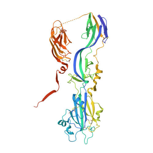 | |
UniProt | |||||
Find proteins for P08563 (Rubella virus (strain M33)) Explore P08563 Go to UniProtKB: P08563 | |||||
Entity Groups | |||||
| Sequence Clusters | 30% Identity50% Identity70% Identity90% Identity95% Identity100% Identity | ||||
| UniProt Group | P08563 | ||||
Sequence AnnotationsExpand | |||||
| |||||
Small Molecules
| Ligands 6 Unique | |||||
|---|---|---|---|---|---|
| ID | Chains | Name / Formula / InChI Key | 2D Diagram | 3D Interactions | |
| NGA Query on NGA | CA [auth B] DA [auth B] P [auth A] Q [auth A] QA [auth C] | 2-acetamido-2-deoxy-beta-D-galactopyranose C8 H15 N O6 OVRNDRQMDRJTHS-JAJWTYFOSA-N |  | ||
| NAG Query on NAG | BA [auth B] D [auth A] EA [auth C] O [auth A] PA [auth C] | 2-acetamido-2-deoxy-beta-D-glucopyranose C8 H15 N O6 OVRNDRQMDRJTHS-FMDGEEDCSA-N |  | ||
| PEG Query on PEG | AA [auth B], N [auth A], NA [auth C], OA [auth C] | DI(HYDROXYETHYL)ETHER C4 H10 O3 MTHSVFCYNBDYFN-UHFFFAOYSA-N |  | ||
| GOL Query on GOL | E [auth A] F [auth A] FA [auth C] G [auth A] GA [auth C] | GLYCEROL C3 H8 O3 PEDCQBHIVMGVHV-UHFFFAOYSA-N |  | ||
| ACT Query on ACT | L [auth A] M [auth A] MA [auth C] W [auth B] X [auth B] | ACETATE ION C2 H3 O2 QTBSBXVTEAMEQO-UHFFFAOYSA-M |  | ||
| NA Query on NA | K [auth A], LA [auth C], V [auth B] | SODIUM ION Na FKNQFGJONOIPTF-UHFFFAOYSA-N |  | ||
Experimental Data & Validation
Experimental Data
- Method: X-RAY DIFFRACTION
- Resolution: 1.80 Å
- R-Value Free: 0.183
- R-Value Work: 0.167
- R-Value Observed: 0.168
- Space Group: P 21 21 21
Unit Cell:
| Length ( Å ) | Angle ( ˚ ) |
|---|---|
| a = 129.98 | α = 90 |
| b = 121.38 | β = 90 |
| c = 126.43 | γ = 90 |
| Software Name | Purpose |
|---|---|
| BUSTER | refinement |
| XDS | data reduction |
| SCALA | data scaling |
| SHARP | phasing |
Entry History
Deposition Data
- Released Date: 2013-01-09 Deposition Author(s): DuBois, R.M., Vaney, M.C., Tortorici, M.A., Al Kurdi, R., Barba-Spaeth, G., Rey, F.A.
Revision History (Full details and data files)
- Version 1.0: 2013-01-09
Type: Initial release - Version 1.1: 2013-01-23
Changes: Database references - Version 1.2: 2020-07-29
Type: Remediation
Reason: Carbohydrate remediation
Changes: Data collection, Derived calculations, Other, Structure summary
