The 1.8 A crystal structure of a statically disordered 17 base-pair RNA duplex: principles of RNA crystal packing and its effect on nucleic acid structure.
Shah, S.A., Brunger, A.T.(1999) J Mol Biol 285: 1577-1588
- PubMed: 9917398
- DOI: https://doi.org/10.1006/jmbi.1998.2385
- Primary Citation of Related Structures:
406D - PubMed Abstract:
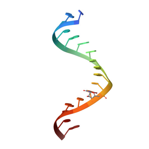
The crystal structure of a 17 base RNA oligomer, r(CACCGGAUG GUUCGGUG), has been solved to a resolution of 1.8 A through a combination of molecular replacement, multiple isomorphous replacement phasing, and analysis of observed intensity distributions. The oligomer, which forms a stem-loop in solution, crystallized as a pseudo-infinite duplex in spacegroup P321. The asymmetric unit of the crystal contains four superimposed orientations of the duplex that are out of register, such that backbones superimpose, but base identity differs. This static disorder was initially discovered by brominating a single residue per strand in the sequence, and observing four peaks per strand in difference maps phased with a native molecular replacement solution. The presence of four superimposed duplex "motifs" related by non-crystallographic hypersymmetry was detected by computing
/2 and Wilson ratios for the observed intensities. The observed ratios matched those produced from calculated intensities of a 4-fold statically disordered model. Multi-conformer simulated annealing refinement against a maximum-likelihood target incorporating experimental phase information was used to refine the 4-fold disordered model to an Rfree and R of 29.35% and 25.5%, respectively. The resulting structure reveals four distinct conformations of the duplex, with an average pairwise backbone rmsd of 2.35 A. The structural differences between the four conformations, which can be attributed to differences in packing environment, highlight the possible influence of crystal packing forces on nucleic acid X-ray structures. Analysis of inter-helical packing between symmetry-related molecules reveals an RNA "zipper" that mediates direct phosphate oxygen-2' hydroxyl interactions between close-packed phosphate-sugar backbones. This may be a general mode for RNA tertiary interaction that does not depend on metal ions or primary sequence.
Organizational Affiliation:
Department of Molecular Biophysics and Biochemistry.