A novel crystal form of pyrrolysyl-tRNA synthetase reveals the pre- and post-aminoacyl-tRNA synthesis conformational states of the adenylate and aminoacyl moieties and an asparagine residue in the catalytic site
Yanagisawa, T., Sumida, T., Ishii, R., Yokoyama, S.(2013) Acta Crystallogr D Biol Crystallogr 69: 5-15
- PubMed: 23275158
- DOI: https://doi.org/10.1107/S0907444912039881
- Primary Citation of Related Structures:
3VQV, 3VQW, 3VQX, 3VQY - PubMed Abstract:
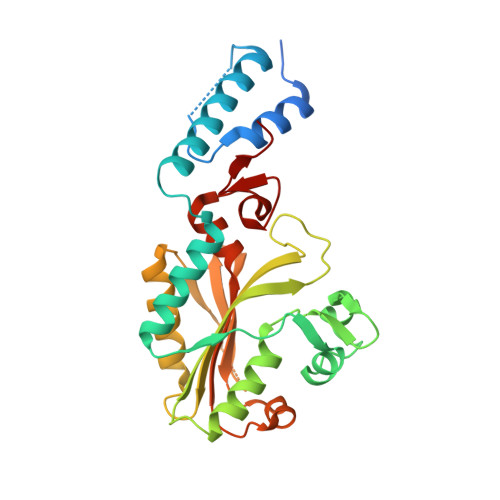
Structures of Methanosarcina mazei pyrrolysyl-tRNA synthetase (PylRS) have been determined in a novel crystal form. The triclinic form crystals contained two PylRS dimers (four monomer molecules) in the asymmetric unit, in which the two subunits in one dimer each bind N(ℇ)-(tert-butyloxycarbonyl)-L-lysyladenylate (BocLys-AMP) and the two subunits in the other dimer each bind AMP. The BocLys-AMP molecules adopt a curved conformation and the C(α) position of BocLys-AMP protrudes from the active site. The β7-β8 hairpin structures in the four PylRS molecules represent distinct conformations of different states of the aminoacyl-tRNA synthesis reaction. Tyr384, at the tip of the β7-β8 hairpin, moves from the edge to the inside of the active-site pocket and adopts multiple conformations in each state. Furthermore, a new crystal structure of the BocLys-AMPPNP-bound form is also reported. The bound BocLys adopts an unusually bent conformation, which differs from the previously reported structure. It is suggested that the present BocLys-AMPPNP-bound, BocLys-AMP-bound and AMP-bound complexes represent the initial binding of an amino acid (or pre-aminoacyl-AMP synthesis), pre-aminoacyl-tRNA synthesis and post-aminoacyl-tRNA synthesis states, respectively. The conformational changes of Asn346 that accompany the aminoacyl-tRNA synthesis reaction have been captured by X-ray crystallographic analyses. The orientation of the Asn346 side chain, which hydrogen-bonds to the carbonyl group of the amino-acid substrate, shifts by a maximum of 85-90° around the C(β) atom.
Organizational Affiliation:
RIKEN Systems and Structural Biology Center, Tsurumi, Yokohama, Japan.


