3CIR

E. coli Quinol fumarate reductase FrdA T234A mutation


Experimental Data Snapshot

  • Method: X-RAY DIFFRACTION
  • Resolution: 3.65 Å
  • R-Value Free: 0.296 
  • R-Value Work: 0.261 

wwPDB Validation   3D Report Full Report


Ligand Structure Quality Assessment 


This is version 1.4 of the entry. See complete history


Literature

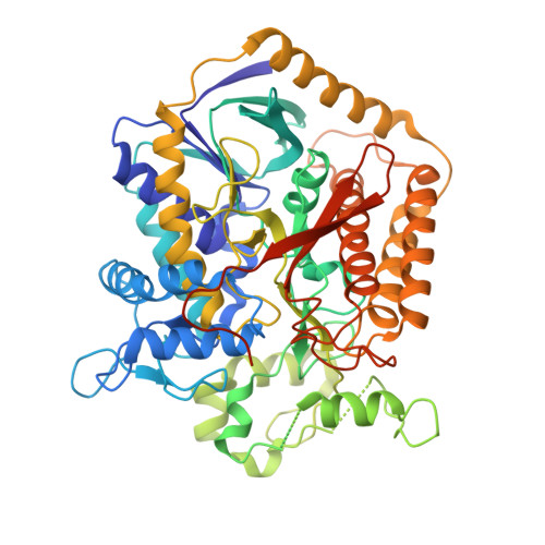
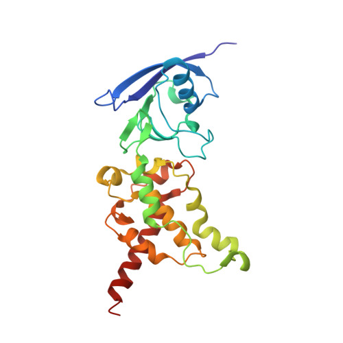
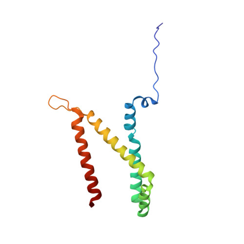
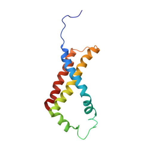
A threonine on the active site loop controls transition state formation in Escherichia coli respiratory complex II.

Tomasiak, T.M.Maklashina, E.Cecchini, G.Iverson, T.M.

(2008) J Biol Chem 283: 15460-15468

  • DOI: https://doi.org/10.1074/jbc.M801372200
  • Primary Citation of Related Structures:  
    3CIR

  • PubMed Abstract: 

    In Escherichia coli, the complex II superfamily members succinate:ubiquinone oxidoreductase (SQR) and quinol:fumarate reductase (QFR) participate in aerobic and anaerobic respiration, respectively. Complex II enzymes catalyze succinate and fumarate interconversion at the interface of two domains of the soluble flavoprotein subunit, the FAD binding domain and the capping domain. An 11-amino acid loop in the capping domain (Thr-A234 to Thr-A244 in quinol:fumarate reductase) begins at the interdomain hinge and covers the active site. Amino acids of this loop interact with both the substrate and a proton shuttle, potentially coordinating substrate binding and the proton shuttle protonation state. To assess the loop's role in catalysis, two threonine residues were mutated to alanine: QFR Thr-A244 (act-T; Thr-A254 in SQR), which hydrogen-bonds to the substrate at the active site, and QFR Thr-A234 (hinge-T; Thr-A244 in SQR), which is located at the hinge and hydrogen-bonds the proton shuttle. Both mutations impair catalysis and decrease substrate binding. The crystal structure of the hinge-T mutation reveals a reorientation between the FAD-binding and capping domains that accompanies proton shuttle alteration. Taken together, hydrogen bonding from act-T to substrate may coordinate with interdomain motions to twist the double bond of fumarate and introduce the strain important for attaining the transition state.


  • Organizational Affiliation

    Department of Pharmacology, Vanderbilt University, Nashville, TN 37232, USA.


Macromolecules
Find similar proteins by:  (by identity cutoff)  |  3D Structure
Entity ID: 1
MoleculeChains Sequence LengthOrganismDetailsImage
Fumarate reductase flavoprotein subunitA,
E [auth M]
602Escherichia coliMutation(s): 1 
Gene Names: FrdA
EC: 1.3.99.1
Membrane Entity: Yes 
UniProt
Find proteins for P00363 (Escherichia coli (strain K12))
Explore P00363 
Go to UniProtKB:  P00363
Entity Groups  
Sequence Clusters30% Identity50% Identity70% Identity90% Identity95% Identity100% Identity
UniProt GroupP00363
Sequence Annotations
Expand
  • Reference Sequence
Find similar proteins by:  (by identity cutoff)  |  3D Structure
Entity ID: 2
MoleculeChains Sequence LengthOrganismDetailsImage
Fumarate reductase iron-sulfur subunitB,
F [auth N]
243Escherichia coliMutation(s): 0 
Gene Names: FrdB
EC: 1.3.99.1
Membrane Entity: Yes 
UniProt
Find proteins for P0AC47 (Escherichia coli (strain K12))
Explore P0AC47 
Go to UniProtKB:  P0AC47
Entity Groups  
Sequence Clusters30% Identity50% Identity70% Identity90% Identity95% Identity100% Identity
UniProt GroupP0AC47
Sequence Annotations
Expand
  • Reference Sequence
Find similar proteins by:  (by identity cutoff)  |  3D Structure
Entity ID: 3
MoleculeChains Sequence LengthOrganismDetailsImage
Fumarate reductase subunit CC,
G [auth O]
130Escherichia coliMutation(s): 0 
Gene Names: FrdC
EC: 1.3.99.1
Membrane Entity: Yes 
UniProt
Find proteins for P0A8Q0 (Escherichia coli (strain K12))
Explore P0A8Q0 
Go to UniProtKB:  P0A8Q0
Entity Groups  
Sequence Clusters30% Identity50% Identity70% Identity90% Identity95% Identity100% Identity
UniProt GroupP0A8Q0
Sequence Annotations
Expand
  • Reference Sequence
Find similar proteins by:  (by identity cutoff)  |  3D Structure
Entity ID: 4
MoleculeChains Sequence LengthOrganismDetailsImage
Fumarate reductase subunit DD,
H [auth P]
119Escherichia coliMutation(s): 0 
Gene Names: FrdD
EC: 1.3.99.1
Membrane Entity: Yes 
UniProt
Find proteins for P0A8Q3 (Escherichia coli (strain K12))
Explore P0A8Q3 
Go to UniProtKB:  P0A8Q3
Entity Groups  
Sequence Clusters30% Identity50% Identity70% Identity90% Identity95% Identity100% Identity
UniProt GroupP0A8Q3
Sequence Annotations
Expand
  • Reference Sequence
Small Molecules
Ligands 4 Unique
IDChains Name / Formula / InChI Key2D Diagram3D Interactions
FAD
Query on FAD

Download Ideal Coordinates CCD File 
I [auth A],
M
FLAVIN-ADENINE DINUCLEOTIDE
C27 H33 N9 O15 P2
VWWQXMAJTJZDQX-UYBVJOGSSA-N
SF4
Query on SF4

Download Ideal Coordinates CCD File 
L [auth B],
P [auth N]
IRON/SULFUR CLUSTER
Fe4 S4
LJBDFODJNLIPKO-UHFFFAOYSA-N
F3S
Query on F3S

Download Ideal Coordinates CCD File 
K [auth B],
O [auth N]
FE3-S4 CLUSTER
Fe3 S4
FCXHZBQOKRZXKS-UHFFFAOYSA-N
FES
Query on FES

Download Ideal Coordinates CCD File 
J [auth B],
N
FE2/S2 (INORGANIC) CLUSTER
Fe2 S2
NIXDOXVAJZFRNF-UHFFFAOYSA-N
Experimental Data & Validation

Experimental Data

  • Method: X-RAY DIFFRACTION
  • Resolution: 3.65 Å
  • R-Value Free: 0.296 
  • R-Value Work: 0.261 
  • Space Group: P 21 21 21
Unit Cell:
Length ( Å )Angle ( ˚ )
a = 96.861α = 90
b = 135.468β = 90
c = 266.024γ = 90
Software Package:
Software NamePurpose
ADSCdata collection
CNSrefinement
DENZOdata reduction
SCALEPACKdata scaling
CNSphasing

Structure Validation

View Full Validation Report


Ligand Structure Quality Assessment 


Entry History 

Deposition Data

Revision History  (Full details and data files)

  • Version 1.0: 2008-04-01
    Type: Initial release
  • Version 1.1: 2011-07-13
    Changes: Version format compliance
  • Version 1.2: 2021-07-28
    Changes: Derived calculations, Refinement description
  • Version 1.3: 2021-10-20
    Changes: Database references
  • Version 1.4: 2023-08-30
    Changes: Data collection, Refinement description