3V7E

Crystal structure of YbxF bound to the SAM-I riboswitch aptamer


Experimental Data Snapshot

  • Method: X-RAY DIFFRACTION
  • Resolution: 2.80 Å
  • R-Value Free: 0.274 
  • R-Value Work: 0.218 
  • R-Value Observed: 0.218 

wwPDB Validation   3D Report Full Report


Ligand Structure Quality Assessment 


This is version 1.2 of the entry. See complete history


Literature

YbxF and YlxQ are bacterial homologs of L7Ae and bind K-turns but not K-loops.

Baird, N.J.Zhang, J.Hamma, T.Ferre-D'Amare, A.R.

(2012) RNA 18: 759-770

  • DOI: https://doi.org/10.1261/rna.031518.111
  • Primary Citation of Related Structures:  
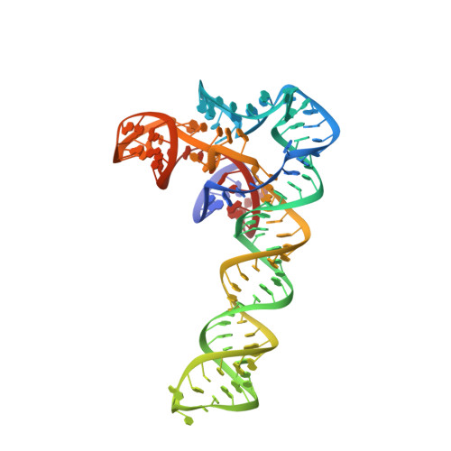
    3V7E, 3V7Q

  • PubMed Abstract: 

    The archaeal protein L7Ae and eukaryotic homologs such as L30e and 15.5kD comprise the best characterized family of K-turn-binding proteins. K-turns are an RNA motif comprised of a bulge flanked by canonical and noncanonical helices. They are widespread in cellular RNAs, including bacterial gene-regulatory RNAs such as the c-di-GMP-II, lysine, and SAM-I riboswitches, and the T-box. The existence in bacteria of K-turn-binding proteins of the L7Ae family has not been proven, although two hypothetical proteins, YbxF and YlxQ, have been proposed to be L7Ae homologs based on sequence conservation. Using purified, recombinant proteins, we show that Bacillus subtilis YbxF and YlxQ bind K-turns (K(d) ~270 nM and ~2300 nM, respectively). Crystallographic structure determination demonstrates that both YbxF and YlxQ adopt the same overall fold as L7Ae. Unlike the latter, neither bacterial protein recognizes K-loops, a structural motif that lacks the canonical helix of the K-turn. This property is shared between the bacterial and eukaryal family members. Comparison of our structure of YbxF in complex with the K-turn of the SAM-I riboswitch and previously determined structures of archaeal and eukaryal homologs bound to RNA indicates that L7Ae approaches the K-turn at a unique angle, which results in a considerably larger RNA-protein interface dominated by interactions with the noncanonical helix of the K-turn. Thus, the inability of the bacterial and eukaryal L7Ae homologs to bind K-loops probably results from their reliance on interactions with the canonical helix. The biological functions of YbxF and YlxQ remain to be determined.


  • Organizational Affiliation

    Laboratory of RNA Biophysics and Cellular Physiology, National Heart, Lung and Blood Institute, Bethesda, Maryland 20892-8012, USA.


Macromolecules

Find similar proteins by:  (by identity cutoff)  |  3D Structure
Entity ID: 1
MoleculeChains Sequence LengthOrganismDetailsImage
Ribosome-associated protein L7Ae-like
A, B
82Bacillus subtilisMutation(s): 0 
Gene Names: BSU01090rplGBybaBybxF
UniProt
Find proteins for P46350 (Bacillus subtilis (strain 168))
Explore P46350 
Go to UniProtKB:  P46350
Entity Groups  
Sequence Clusters30% Identity50% Identity70% Identity90% Identity95% Identity100% Identity
UniProt GroupP46350
Sequence Annotations
Expand
  • Reference Sequence
Find similar nucleic acids by:  (by identity cutoff)  |  3D Structure
Entity ID: 2
MoleculeChains LengthOrganismImage
SAM-I riboswitch aptamer with an engineered helix P3
C, D
126Caldanaerobacter subterraneus subsp. tengcongensis
Sequence Annotations
Expand
  • Reference Sequence
Small Molecules
Ligands 3 Unique
IDChains Name / Formula / InChI Key2D Diagram3D Interactions
SAM
Query on SAM

Download Ideal Coordinates CCD File 
KA [auth D],
Q [auth C]
S-ADENOSYLMETHIONINE
C15 H22 N6 O5 S
MEFKEPWMEQBLKI-FCKMPRQPSA-N
NCO
Query on NCO

Download Ideal Coordinates CCD File 
E [auth C]
EA [auth D]
F [auth C]
FA [auth D]
G [auth C]
E [auth C],
EA [auth D],
F [auth C],
FA [auth D],
G [auth C],
GA [auth D],
H [auth C],
HA [auth D],
I [auth C],
IA [auth D],
J [auth C],
JA [auth D],
K [auth C],
L [auth C],
M [auth C],
N [auth C],
O [auth C],
P [auth C]
COBALT HEXAMMINE(III)
Co H18 N6
DYLMFCCYOUSRTK-UHFFFAOYSA-N
MG
Query on MG

Download Ideal Coordinates CCD File 
AA [auth C]
BA [auth C]
CA [auth C]
DA [auth C]
LA [auth D]
AA [auth C],
BA [auth C],
CA [auth C],
DA [auth C],
LA [auth D],
MA [auth D],
NA [auth D],
OA [auth D],
PA [auth D],
QA [auth D],
R [auth C],
RA [auth D],
S [auth C],
SA [auth D],
T [auth C],
TA [auth D],
U [auth C],
V [auth C],
W [auth C],
X [auth C],
Y [auth C],
Z [auth C]
MAGNESIUM ION
Mg
JLVVSXFLKOJNIY-UHFFFAOYSA-N
Experimental Data & Validation

Experimental Data

  • Method: X-RAY DIFFRACTION
  • Resolution: 2.80 Å
  • R-Value Free: 0.274 
  • R-Value Work: 0.218 
  • R-Value Observed: 0.218 
  • Space Group: C 1 2 1
Unit Cell:
Length ( Å )Angle ( ˚ )
a = 191.762α = 90
b = 54.305β = 116.56
c = 106.366γ = 90
Software Package:
Software NamePurpose
ALSdata collection
PHASERphasing
CNSrefinement
HKL-2000data reduction
HKL-2000data scaling

Structure Validation

View Full Validation Report


Ligand Structure Quality Assessment 


Entry History 

Deposition Data

Revision History  (Full details and data files)

  • Version 1.0: 2012-03-07
    Type: Initial release
  • Version 1.1: 2012-04-18
    Changes: Database references
  • Version 1.2: 2023-09-13
    Changes: Data collection, Database references, Derived calculations, Refinement description