3UZT

Structure of the C13.18 RNA Aptamer in Complex with G Protein-Coupled Receptor Kinase 2


Experimental Data Snapshot

  • Method: X-RAY DIFFRACTION
  • Resolution: 3.51 Å
  • R-Value Free: 0.317 
  • R-Value Work: 0.216 
  • R-Value Observed: 0.220 

wwPDB Validation   3D Report Full Report


This is version 1.2 of the entry. See complete history


Literature

Molecular mechanism for inhibition of g protein-coupled receptor kinase 2 by a selective RNA aptamer.

Tesmer, V.M.Lennarz, S.Mayer, G.Tesmer, J.J.

(2012) Structure 20: 1300-1309

  • DOI: https://doi.org/10.1016/j.str.2012.05.002
  • Primary Citation of Related Structures:  
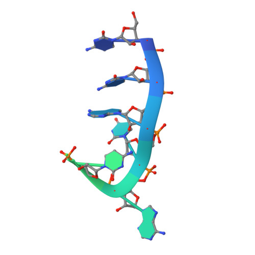
    3UZS, 3UZT

  • PubMed Abstract: 

    Cardiovascular homeostasis is maintained in part by the rapid desensitization of activated heptahelical receptors that have been phosphorylated by G protein-coupled receptor kinase 2 (GRK2). However, during chronic heart failure GRK2 is upregulated and believed to contribute to disease progression. We have determined crystallographic structures of GRK2 bound to an RNA aptamer that potently and selectively inhibits kinase activity. Key to the mechanism of inhibition is the positioning of an adenine nucleotide into the ATP-binding pocket and interactions with the basic αF-αG loop region of the GRK2 kinase domain. Constraints imposed on the RNA by the terminal stem of the aptamer also play a role. These results highlight how a high-affinity aptamer can be used to selectively trap a novel conformational state of a protein kinase.


  • Organizational Affiliation

    Life Sciences Institute, University of Michigan, 210 Washtenaw Avenue, Ann Arbor, MI 48109-2216, USA. tesmerjj@umich.edu


Macromolecules

Find similar proteins by:  (by identity cutoff)  |  3D Structure
Entity ID: 1
MoleculeChains Sequence LengthOrganismDetailsImage
Beta-adrenergic receptor kinase 1689Bos taurusMutation(s): 1 
Gene Names: ADRBK1GRK2
EC: 2.7.11.15
UniProt
Find proteins for P21146 (Bos taurus)
Explore P21146 
Go to UniProtKB:  P21146
Entity Groups  
Sequence Clusters30% Identity50% Identity70% Identity90% Identity95% Identity100% Identity
UniProt GroupP21146
Sequence Annotations
Expand
  • Reference Sequence

Find similar nucleic acids by:  Sequence   |   3D Structure  

Entity ID: 2
MoleculeChains LengthOrganismImage
C13.18 RNA Aptamer18N/A
Sequence Annotations
Expand
  • Reference Sequence
Small Molecules
Ligands 1 Unique
IDChains Name / Formula / InChI Key2D Diagram3D Interactions
MG
Query on MG

Download Ideal Coordinates CCD File 
C [auth A]MAGNESIUM ION
Mg
JLVVSXFLKOJNIY-UHFFFAOYSA-N
Experimental Data & Validation

Experimental Data

  • Method: X-RAY DIFFRACTION
  • Resolution: 3.51 Å
  • R-Value Free: 0.317 
  • R-Value Work: 0.216 
  • R-Value Observed: 0.220 
  • Space Group: P 21 21 2
Unit Cell:
Length ( Å )Angle ( ˚ )
a = 113.346α = 90
b = 139.719β = 90
c = 60.949γ = 90
Software Package:
Software NamePurpose
MAR345dtbdata collection
PHASERphasing
REFMACrefinement
HKL-2000data reduction
SCALEPACKdata scaling

Structure Validation

View Full Validation Report


Entry History 

Deposition Data

Revision History  (Full details and data files)

  • Version 1.0: 2012-07-11
    Type: Initial release
  • Version 1.1: 2012-08-29
    Changes: Database references
  • Version 1.2: 2023-09-13
    Changes: Data collection, Database references, Derived calculations, Refinement description