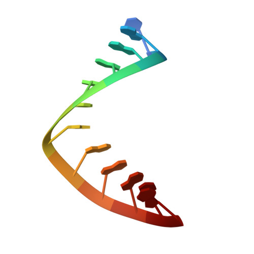
Structure of an RNA/DNA dodecamer corresponding to the HIV-1 polypurine tract at 1.6 Angstrom resolution
Drozdzal, P., Michalska, K., Kierzek, R., Lomozik, L., Jaskolski, M.(2012) Acta Crystallogr D Biol Crystallogr 68: 169-175
- PubMed: 22281746
- DOI: https://doi.org/10.1107/S0907444911053327
- Primary Citation of Related Structures:
3SSF - PubMed Abstract:
The crystal structure of an RNA/DNA hybrid dodecamer, r(5'-uaaaagaaaagg):d(5'-CCTTTTCTTTTA), which contains three-quarters of the polypurine tract (PPT) sequence of the HIV RNA genome is reported. The hybrid structure was determined at 1.6 Å resolution and was found to have the A-form conformation. However, the presence of alternate conformations along the RNA template strand indicated increased flexibility of the PPT sequence. Two segments (at nucleotides 1-2 and 6-8) of the RNA chain have two conformations exhibiting differences in torsion and pseudorotation angles. For conformation I((1-2), (6-8)), 25% of the RNA sugars have the C2'-exo pucker and the rest have the expected C3'-endo pucker. The II(1-2) and II(6-8) conformations of the RNA strand have one sugar with the C2'-exo pucker. None of the ribose rings exist in the C2'-endo form, in contrast to a previous report which postulated a C2'-endo ribose as a key structural element of the PPT. The widths of the minor groove for conformations I((1-2), (6-8)) and II((1-2), (6-8)) of the RNA strand are 9.2-10.5 and 9.4-10.7 Å, respectively. Both ranges are very close to the intervals accepted for A-form RNA duplexes. On the opposing DNA primer strand most of the sugars are C3'-endo, except for the 3'-terminal sugars, which are C2'-endo (T22) or O4'-endo (T23 and A24). The duplex includes a noncanonical u1(anti)·A24(syn) base interaction with only one hydrogen bond between the bases. This noncanonical base interaction at the 5'-end of the template distorts the values of the helical parameters of the adjacent base pair.
Organizational Affiliation:
Faculty of Chemistry, A. Mickiewicz University, 60-780 Poznan, Poland.