3SKR

Crystal structure of the 2'- Deoxyguanosine riboswitch bound to 2'- Deoxyguanosine, cobalt Hexammine soak


Experimental Data Snapshot

  • Method: X-RAY DIFFRACTION
  • Resolution: 3.10 Å
  • R-Value Free: 0.299 
  • R-Value Work: 0.236 
  • R-Value Observed: 0.242 

wwPDB Validation   3D Report Full Report


Ligand Structure Quality Assessment 


This is version 1.3 of the entry. See complete history


Literature

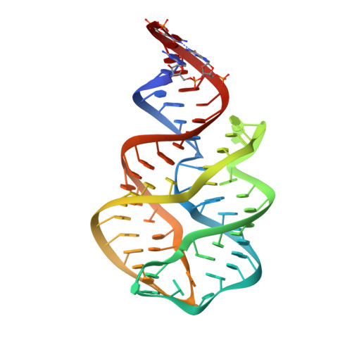
Structural principles of nucleoside selectivity in a 2'-deoxyguanosine riboswitch.

Pikovskaya, O.Polonskaia, A.Patel, D.J.Serganov, A.

(2011) Nat Chem Biol 7: 748-755

  • DOI: https://doi.org/10.1038/nchembio.631
  • Primary Citation of Related Structures:  
    3SKI, 3SKL, 3SKR, 3SKT, 3SKW, 3SKZ, 3SLM, 3SLQ

  • PubMed Abstract: 

    Purine riboswitches have an essential role in genetic regulation of bacterial metabolism. This family includes the 2'-deoxyguanosine (dG) riboswitch, which is involved in feedback control of deoxyguanosine biosynthesis. To understand the principles that define dG selectivity, we determined crystal structures of the natural Mesoplasma florum riboswitch bound to cognate dG as well as to noncognate guanosine, deoxyguanosine monophosphate and guanosine monophosphate. Comparison with related purine riboswitch structures reveals that the dG riboswitch achieves its specificity through modification of key interactions involving the nucleobase and rearrangement of the ligand-binding pocket to accommodate the additional sugar moiety. In addition, we observe new conformational changes beyond the junctional binding pocket extending as far as peripheral loop-loop interactions. It appears that re-engineering riboswitch scaffolds will require consideration of selectivity features dispersed throughout the riboswitch tertiary fold, and structure-guided drug design efforts targeted to junctional RNA scaffolds need to be addressed within such an expanded framework.


  • Organizational Affiliation

    Structural Biology Program, Memorial Sloan-Kettering Cancer Center, New York, New York, USA.


Macromolecules
Find similar nucleic acids by:  (by identity cutoff)  |  3D Structure
Entity ID: 1
MoleculeChains LengthOrganismImage
RNA (66-MER)
A, B
66N/A
Sequence Annotations
Expand
  • Reference Sequence
Small Molecules
Ligands 3 Unique
IDChains Name / Formula / InChI Key2D Diagram3D Interactions
GNG
Query on GNG

Download Ideal Coordinates CCD File 
C [auth A],
L [auth B]
2'-DEOXY-GUANOSINE
C10 H13 N5 O4
YKBGVTZYEHREMT-KVQBGUIXSA-N
NCO
Query on NCO

Download Ideal Coordinates CCD File 
D [auth A]
E [auth A]
F [auth A]
G [auth A]
H [auth A]
D [auth A],
E [auth A],
F [auth A],
G [auth A],
H [auth A],
M [auth B],
N [auth B],
O [auth B],
P [auth B]
COBALT HEXAMMINE(III)
Co H18 N6
DYLMFCCYOUSRTK-UHFFFAOYSA-N
MG
Query on MG

Download Ideal Coordinates CCD File 
I [auth A],
J [auth A],
K [auth A],
Q [auth B]
MAGNESIUM ION
Mg
JLVVSXFLKOJNIY-UHFFFAOYSA-N
Experimental Data & Validation

Experimental Data

  • Method: X-RAY DIFFRACTION
  • Resolution: 3.10 Å
  • R-Value Free: 0.299 
  • R-Value Work: 0.236 
  • R-Value Observed: 0.242 
  • Space Group: C 1 2 1
Unit Cell:
Length ( Å )Angle ( ˚ )
a = 98.53α = 90
b = 35.018β = 92.18
c = 110.761γ = 90
Software Package:
Software NamePurpose
CBASSdata collection
PHASERphasing
REFMACrefinement
HKL-2000data reduction
HKL-2000data scaling

Structure Validation

View Full Validation Report


Ligand Structure Quality Assessment 


Entry History 

Deposition Data

Revision History  (Full details and data files)

  • Version 1.0: 2011-08-17
    Type: Initial release
  • Version 1.1: 2011-10-26
    Changes: Database references
  • Version 1.2: 2024-02-28
    Changes: Data collection, Database references, Derived calculations
  • Version 1.3: 2024-03-13
    Changes: Source and taxonomy, Structure summary