3P59

First Crystal Structure of a RNA Nanosquare


Experimental Data Snapshot

  • Method: X-RAY DIFFRACTION
  • Resolution: 2.18 Å
  • R-Value Free: 0.192 
  • R-Value Work: 0.156 
  • R-Value Observed: 0.157 

wwPDB Validation   3D Report Full Report


This is version 1.2 of the entry. See complete history


Literature

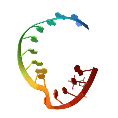
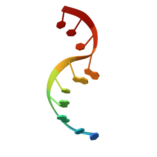
Self-assembling RNA square.

Dibrov, S.M.McLean, J.Parsons, J.Hermann, T.

(2011) Proc Natl Acad Sci U S A 108: 6405-6408

  • DOI: https://doi.org/10.1073/pnas.1017999108
  • Primary Citation of Related Structures:  
    3P59

  • PubMed Abstract: 

    The three-dimensional structures of noncoding RNA molecules reveal recurring architectural motifs that have been exploited for the design of artificial RNA nanomaterials. Programmed assembly of RNA nanoobjects from autonomously folding tetraloop-receptor complexes as well as junction motifs has been achieved previously through sequence-directed hybridization of complex sets of long oligonucleotides. Due to size and complexity, structural characterization of artificial RNA nanoobjects has been limited to low-resolution microscopy studies. Here we present the design, construction, and crystal structure determination at 2.2 Å of the smallest yet square-shaped nanoobject made entirely of double-stranded RNA. The RNA square is comprised of 100 residues and self-assembles from four copies each of two oligonucleotides of 10 and 15 bases length. Despite the high symmetry on the level of secondary structure, the three-dimensional architecture of the square is asymmetric, with all four corners adopting distinct folding patterns. We demonstrate the programmed self-assembly of RNA squares from complex mixtures of corner units and establish a concept to exploit the RNA square as a combinatorial nanoscale platform.


  • Organizational Affiliation

    Department of Chemistry and Biochemistry, University of California at San Diego, 9500 Gilman Drive, La Jolla, CA 92093, USA.


Macromolecules

Find similar nucleic acids by:  Sequence   |   3D Structure  

Entity ID: 1
MoleculeChains LengthOrganismImage
RNA (5'-R(*CP*CP*GP*GP*AP*GP*GP*AP*AP*CP*UP*AP*CP*(5BU)P*G)-3')
A, C, E, G
15N/A
Sequence Annotations
Expand
  • Reference Sequence

Find similar nucleic acids by:  Sequence   |   3D Structure  

Entity ID: 2
MoleculeChains LengthOrganismImage
RNA (5'-R(*CP*CP*GP*GP*CP*AP*GP*CP*CP*U)-3')
B, D, F, H
10N/A
Sequence Annotations
Expand
  • Reference Sequence
Small Molecules
Ligands 2 Unique
IDChains Name / Formula / InChI Key2D Diagram3D Interactions
NCO
Query on NCO

Download Ideal Coordinates CCD File 
I [auth A]
K [auth B]
L [auth C]
M [auth C]
N [auth D]
I [auth A],
K [auth B],
L [auth C],
M [auth C],
N [auth D],
O [auth E],
Q [auth F],
R [auth G],
S [auth G],
T [auth H]
COBALT HEXAMMINE(III)
Co H18 N6
DYLMFCCYOUSRTK-UHFFFAOYSA-N
MG
Query on MG

Download Ideal Coordinates CCD File 
J [auth A],
P [auth E]
MAGNESIUM ION
Mg
JLVVSXFLKOJNIY-UHFFFAOYSA-N
Experimental Data & Validation

Experimental Data

  • Method: X-RAY DIFFRACTION
  • Resolution: 2.18 Å
  • R-Value Free: 0.192 
  • R-Value Work: 0.156 
  • R-Value Observed: 0.157 
  • Space Group: P 31 2 1
Unit Cell:
Length ( Å )Angle ( ˚ )
a = 62.378α = 90
b = 62.378β = 90
c = 126.268γ = 120
Software Package:
Software NamePurpose
HKL-2000data collection
PHENIXmodel building
PHENIXrefinement
HKL-2000data reduction
HKL-2000data scaling
PHENIXphasing

Structure Validation

View Full Validation Report


Entry History 

Deposition Data

Revision History  (Full details and data files)

  • Version 1.0: 2011-04-06
    Type: Initial release
  • Version 1.1: 2011-07-13
    Changes: Version format compliance
  • Version 1.2: 2024-02-21
    Changes: Data collection, Database references, Derived calculations