3N2J

Azurin H117G, oxidized form


Experimental Data Snapshot

  • Method: X-RAY DIFFRACTION
  • Resolution: 1.35 Å
  • R-Value Free: 0.202 
  • R-Value Work: 0.172 
  • R-Value Observed: 0.174 

wwPDB Validation   3D Report Full Report


This is version 1.4 of the entry. See complete history


Literature

Probing the reactivity of different forms of azurin by flavin photoreduction.

Alagaratnam, S.Meeuwenoord, N.J.Navarro, J.A.Hervas, M.De la Rosa, M.A.Hoffmann, M.Einsle, O.Ubbink, M.Canters, G.W.

(2011) FEBS J 278: 1506-1521

  • DOI: https://doi.org/10.1111/j.1742-4658.2011.08067.x
  • Primary Citation of Related Structures:  
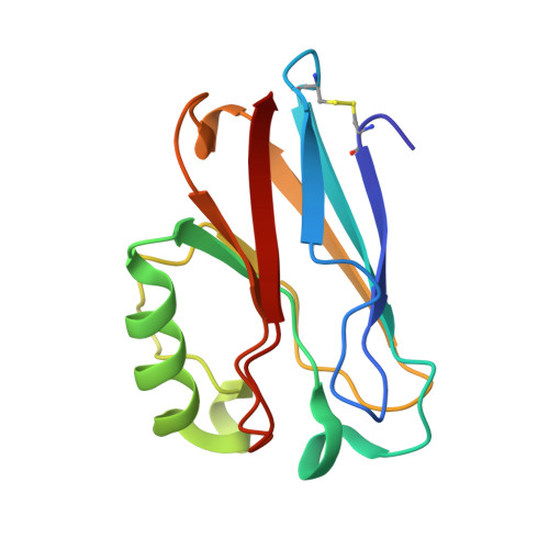
    3N2J

  • PubMed Abstract: 

    The reactivity of a variant of the blue copper protein, azurin from Pseudomonas aeruginosa, was investigated with laser flash photolysis and compared with the reactivity of the wild-type (WT) protein. The variant was obtained by changing the Cu ligating His117 for a glycine. The mutation creates a gap in the ligand shell of the Cu that can be filled with external ligands or water molecules. The crystal structure of the H117G variant is reported. It shows that the immediate surrounding of the Cu site in the variant exhibits less rigidity than in the WT protein and that the loop containing the Cu ligands Cys112, His117 and Met121 in the WT protein has gained flexibility in the H117G variant. Flash photolysis experiments were performed with 5-deazariboflavin and 8α-imidazolyl-(N-propylyl)-amino riboflavin as electron donors to probe the reactivity of WT and H117G azurin, and of H117G azurin for which the gap in the Cu co-ordination shell was filled with imidazole. 8α-Imidazolyl-(N-propylyl)-amino riboflavin appears one to two orders less efficient as a photo-flash reductant than 5-deazariboflavin. The reactivity of the H117G variant in the absence of external ligands appears to be 2.5-fold lower than the WT reactivity (second-order rate constants of 51 ± 2 × 10(7) m(-1) ·s(-1) versus 21 ± 1 × 10(7) m(-1) ·s(-1) ), whereas the addition of imidazole restores reactivity to above the WT level (71 ± 4 × 10(7) m(-1) ·s(-1) ). The differences are discussed in terms of structural modifications and changes in reorganizational energy and electronic coupling. Database Structural data are available in the Protein Data Bank under the accession number 3N2J.


  • Organizational Affiliation

    Gorlaeus Laboratories, Leiden Institute of Chemistry, Leiden University, Leiden, The Netherlands.


Macromolecules
Find similar proteins by:  (by identity cutoff)  |  3D Structure
Entity ID: 1
MoleculeChains Sequence LengthOrganismDetailsImage
Azurin
A, B, C, D, E
A, B, C, D, E, F, G, H, I, J, K, L
128Pseudomonas aeruginosaMutation(s): 1 
Gene Names: azuPA4922
UniProt
Find proteins for P00282 (Pseudomonas aeruginosa (strain ATCC 15692 / DSM 22644 / CIP 104116 / JCM 14847 / LMG 12228 / 1C / PRS 101 / PAO1))
Explore P00282 
Go to UniProtKB:  P00282
Entity Groups  
Sequence Clusters30% Identity50% Identity70% Identity90% Identity95% Identity100% Identity
UniProt GroupP00282
Sequence Annotations
Expand
  • Reference Sequence
Small Molecules
Ligands 1 Unique
IDChains Name / Formula / InChI Key2D Diagram3D Interactions
CU
Query on CU

Download Ideal Coordinates CCD File 
AA [auth H]
BA [auth H]
CA [auth I]
DA [auth I]
EA [auth J]
AA [auth H],
BA [auth H],
CA [auth I],
DA [auth I],
EA [auth J],
FA [auth J],
GA [auth K],
HA [auth K],
IA [auth L],
JA [auth L],
M [auth A],
N [auth A],
O [auth B],
P [auth B],
Q [auth C],
R [auth C],
S [auth D],
T [auth D],
U [auth E],
V [auth E],
W [auth F],
X [auth F],
Y [auth G],
Z [auth G]
COPPER (II) ION
Cu
JPVYNHNXODAKFH-UHFFFAOYSA-N
Experimental Data & Validation

Experimental Data

  • Method: X-RAY DIFFRACTION
  • Resolution: 1.35 Å
  • R-Value Free: 0.202 
  • R-Value Work: 0.172 
  • R-Value Observed: 0.174 
  • Space Group: P 1 21 1
Unit Cell:
Length ( Å )Angle ( ˚ )
a = 47.545α = 90
b = 170.008β = 89.98
c = 85.737γ = 90
Software Package:
Software NamePurpose
MAR345data collection
MOLREPphasing
BUSTERrefinement
DENZOdata reduction
SCALEPACKdata scaling

Structure Validation

View Full Validation Report


Entry History 

Deposition Data

Revision History  (Full details and data files)

  • Version 1.0: 2011-04-06
    Type: Initial release
  • Version 1.1: 2011-07-13
    Changes: Version format compliance
  • Version 1.2: 2011-09-14
    Changes: Database references
  • Version 1.3: 2017-11-08
    Changes: Refinement description
  • Version 1.4: 2023-09-06
    Changes: Data collection, Database references, Derived calculations, Refinement description