3MEI

Regulatory motif from the thymidylate synthase mRNA


Experimental Data Snapshot

  • Method: X-RAY DIFFRACTION
  • Resolution: 1.97 Å
  • R-Value Free: 0.238 
  • R-Value Work: 0.176 
  • R-Value Observed: 0.179 

wwPDB Validation   3D Report Full Report


This is version 1.5 of the entry. See complete history


Literature

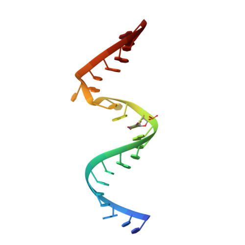
Structure of an RNA dimer of a regulatory element from human thymidylate synthase mRNA.

Dibrov, S.McLean, J.Hermann, T.

(2011) Acta Crystallogr D Biol Crystallogr 67: 97-104

  • DOI: https://doi.org/10.1107/S0907444910050900
  • Primary Citation of Related Structures:  
    3MEI

  • PubMed Abstract: 

    A sequence around the start codon of the mRNA of human thymidylate synthase (TS) folds into a secondary-structure motif in which the initiation site is sequestered in a metastable hairpin. Binding of the protein to its own mRNA at the hairpin prevents the production of TS through a translation-repression feedback mechanism. Stabilization of the mRNA hairpin by other ligands has been proposed as a strategy to reduce TS levels in anticancer therapy. Rapidly proliferating cells require high TS activity to maintain the production of thymidine as a building block for DNA synthesis. The crystal structure of a model oligonucleotide (TS1) that represents the TS-binding site of the mRNA has been determined. While fluorescence studies showed that the TS1 RNA preferentially adopts a hairpin structure in solution, even at high RNA concentrations, an asymmetric dimer of two hybridized TS1 strands was obtained in the crystal. The TS1 dimer contains an unusual S-turn motif that also occurs in the `off' state of the human ribosomal decoding site RNA.


  • Organizational Affiliation

    Department of Chemistry and Biochemistry, University of California, San Diego, 9500 Gilman Drive, La Jolla, CA 92093, USA.


Macromolecules

Find similar nucleic acids by:  Sequence   |   3D Structure  

Entity ID: 1
MoleculeChains LengthOrganismImage
RNA (5'-R(*CP*CP*GP*CP*CP*GP*CP*GP*CP*CP*AP*(5BU)P*GP*CP*CP*UP*GP*UP*GP*GP*CP*GP*G)-3')
A, B
23N/A
Sequence Annotations
Expand
  • Reference Sequence
Experimental Data & Validation

Experimental Data

  • Method: X-RAY DIFFRACTION
  • Resolution: 1.97 Å
  • R-Value Free: 0.238 
  • R-Value Work: 0.176 
  • R-Value Observed: 0.179 
  • Space Group: P 1 21 1
Unit Cell:
Length ( Å )Angle ( ˚ )
a = 32.928α = 90
b = 38.564β = 100.1
c = 41.985γ = 90
Software Package:
Software NamePurpose
PHENIXrefinement
SCALEPACKdata scaling
CNSphasing
CNSrefinement
DENZOdata reduction
PDB_EXTRACTdata extraction
MAR345dtbdata collection
HKL-2000data reduction
HKL-2000data scaling

Structure Validation

View Full Validation Report


Entry History 

Deposition Data

Revision History  (Full details and data files)

  • Version 1.0: 2011-01-26
    Type: Initial release
  • Version 1.1: 2011-07-13
    Changes: Version format compliance
  • Version 1.2: 2012-03-21
    Changes: Database references
  • Version 1.3: 2017-11-08
    Changes: Refinement description
  • Version 1.4: 2019-07-17
    Changes: Data collection, Derived calculations, Refinement description
  • Version 1.5: 2024-02-21
    Changes: Data collection, Database references, Derived calculations