3LOA

Crystal Structure Analysis of the RNA construct with two adjacent ligand binding sites of helix h44 in 16S ribosomal RNA

  • Classification: RNA
  • Mutation(s): No 

  • Deposited: 2010-02-03 Released: 2010-08-04 
  • Deposition Author(s): Dibrov, S.

Experimental Data Snapshot

  • Method: X-RAY DIFFRACTION
  • Resolution: 2.30 Å
  • R-Value Free: 0.231 
  • R-Value Work: 0.191 
  • R-Value Observed: 0.192 

wwPDB Validation   3D Report Full Report


This is version 1.3 of the entry. See complete history


Literature

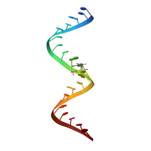
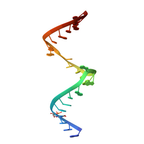
A model for the study of ligand binding to the ribosomal RNA helix h44.

Dibrov, S.M.Parsons, J.Hermann, T.

(2010) Nucleic Acids Res 38: 4458-4465

  • DOI: https://doi.org/10.1093/nar/gkq159
  • Primary Citation of Related Structures:  
    3LOA

  • PubMed Abstract: 

    Oligonucleotide models of ribosomal RNA domains are powerful tools to study the binding and molecular recognition of antibiotics that interfere with bacterial translation. Techniques such as selective chemical modification, fluorescence labeling and mutations are cumbersome for the whole ribosome but readily applicable to model RNAs, which are readily crystallized and often give rise to higher resolution crystal structures suitable for detailed analysis of ligand-RNA interactions. Here, we have investigated the HX RNA construct which contains two adjacent ligand binding regions of helix h44 in 16S ribosomal RNA. High-resolution crystal structure analysis confirmed that the HX RNA is a faithful structural model of the ribosomal target. Solution studies showed that HX RNA carrying a fluorescent 2-aminopurine modification provides a model system that can be used to monitor ligand binding to both the ribosomal decoding site and, through an indirect effect, the hygromycin B interaction region.


  • Organizational Affiliation

    Department of Chemistry and Biochemistry, University of California, San Diego, 9500 Gilman Drive, La Jolla, CA 92093, USA.


Macromolecules

Find similar nucleic acids by:  Sequence   |   3D Structure  

Entity ID: 1
MoleculeChains LengthOrganismImage
5'-R(*CP*CP*GP*CP*GP*CP*CP*CP*GP*(5BU)P*CP*AP*CP*AP*CP*CP*AP*CP*CP*CP*G)-3'21N/A
Sequence Annotations
Expand
  • Reference Sequence

Find similar nucleic acids by:  Sequence   |   3D Structure  

Entity ID: 2
MoleculeChains LengthOrganismImage
5'-R(*GP*GP*GP*(5BU)P*GP*GP*UP*GP*AP*AP*GP*UP*CP*GP*UP*AP*AP*CP*GP*CP*GP*GP*C)-3'23N/A
Sequence Annotations
Expand
  • Reference Sequence
Experimental Data & Validation

Experimental Data

  • Method: X-RAY DIFFRACTION
  • Resolution: 2.30 Å
  • R-Value Free: 0.231 
  • R-Value Work: 0.191 
  • R-Value Observed: 0.192 
  • Space Group: P 64
Unit Cell:
Length ( Å )Angle ( ˚ )
a = 57.455α = 90
b = 57.455β = 90
c = 58.433γ = 120
Software Package:
Software NamePurpose
PHENIXrefinement
CNSrefinement
HKL-2000data collection
HKL-2000data reduction
HKL-2000data scaling
CNSphasing

Structure Validation

View Full Validation Report


Entry History 

Deposition Data

  • Released Date: 2010-08-04 
  • Deposition Author(s): Dibrov, S.

Revision History  (Full details and data files)

  • Version 1.0: 2010-08-04
    Type: Initial release
  • Version 1.1: 2011-07-13
    Changes: Version format compliance
  • Version 1.2: 2019-07-17
    Changes: Data collection, Derived calculations, Refinement description
  • Version 1.3: 2024-02-21
    Changes: Data collection, Database references, Derived calculations