3G6O

Crystal structure of P. aeruginosa bacteriophytochrome PaBphP photosensory core domain mutant Q188L


Experimental Data Snapshot

  • Method: X-RAY DIFFRACTION
  • Resolution: 2.85 Å
  • R-Value Free: 0.298 
  • R-Value Work: 0.229 
  • R-Value Observed: 0.232 

wwPDB Validation   3D Report Full Report


Ligand Structure Quality Assessment 


This is version 1.2 of the entry. See complete history


Literature

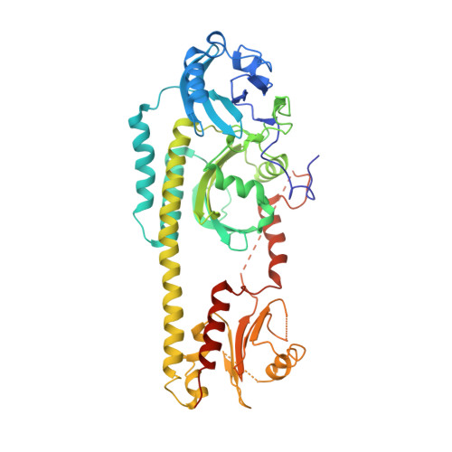
Conformational differences between the Pfr and Pr states in Pseudomonas aeruginosa bacteriophytochrome

Yang, X.Kuk, J.Moffat, K.

(2009) Proc Natl Acad Sci U S A 106: 15639-15644

  • DOI: https://doi.org/10.1073/pnas.0902178106
  • Primary Citation of Related Structures:  
    3G6O, 3IBR

  • PubMed Abstract: 

    Phytochromes are red-light photoreceptors that regulate light responses in plants, fungi, and bacteria by means of reversible photoconversion between red (Pr) and far-red (Pfr) light-absorbing states. Here, we report the crystal structure of the Q188L mutant of Pseudomonas aeruginosa bacteriophytochrome (PaBphP) photosensory core module, which exhibits altered photoconversion behavior and different crystal packing from wild type. We observe two distinct chromophore conformations in the Q188L crystal structure that we identify with the Pfr and Pr states. The Pr/Pfr compositions, varying from crystal to crystal, seem to correlate with light conditions under which the Q188L crystals are cryoprotected. We also compare all known Pr and Pfr structures. Using site-directed mutagenesis, we identify residues that are involved in stabilizing the 15Ea (Pfr) and 15Za (Pr) configurations of the biliverdin chromophore. Specifically, Ser-261 appears to be essential to form a stable Pr state in PaBphP, possibly by means of its interaction with the propionate group of ring C. We propose a "flip-and-rotate" model that summarizes the major conformational differences between the Pr and Pfr states of the chromophore and its binding pocket.


  • Organizational Affiliation

    Department of Biochemistry and Molecular Biology, University of Chicago, 929 East 57th Street, Chicago, IL 60637, USA. xiaojingyang@uchicago.edu


Macromolecules
Find similar proteins by:  (by identity cutoff)  |  3D Structure
Entity ID: 1
MoleculeChains Sequence LengthOrganismDetailsImage
Bacteriophytochrome
A, B
505Pseudomonas aeruginosaMutation(s): 1 
Gene Names: bphPPA4117
EC: 2.7.13.3
UniProt
Find proteins for Q9HWR3 (Pseudomonas aeruginosa (strain ATCC 15692 / DSM 22644 / CIP 104116 / JCM 14847 / LMG 12228 / 1C / PRS 101 / PAO1))
Explore Q9HWR3 
Go to UniProtKB:  Q9HWR3
Entity Groups  
Sequence Clusters30% Identity50% Identity70% Identity90% Identity95% Identity100% Identity
UniProt GroupQ9HWR3
Sequence Annotations
Expand
  • Reference Sequence
Small Molecules
Experimental Data & Validation

Experimental Data

  • Method: X-RAY DIFFRACTION
  • Resolution: 2.85 Å
  • R-Value Free: 0.298 
  • R-Value Work: 0.229 
  • R-Value Observed: 0.232 
  • Space Group: P 65
Unit Cell:
Length ( Å )Angle ( ˚ )
a = 109.786α = 90
b = 109.786β = 90
c = 189.113γ = 120
Software Package:
Software NamePurpose
BioCARSdata collection
SHARPphasing
PHENIXrefinement
HKL-2000data reduction
HKL-2000data scaling

Structure Validation

View Full Validation Report


Ligand Structure Quality Assessment 


Entry History 

Deposition Data

Revision History  (Full details and data files)

  • Version 1.0: 2009-09-22
    Type: Initial release
  • Version 1.1: 2011-07-13
    Changes: Version format compliance
  • Version 1.2: 2021-10-20
    Changes: Database references, Derived calculations