3F73

Alignment of guide-target seed duplex within an argonaute silencing complex


Experimental Data Snapshot

  • Method: X-RAY DIFFRACTION
  • Resolution: 3.00 Å
  • R-Value Free: 0.282 
  • R-Value Work: 0.221 
  • R-Value Observed: 0.221 

wwPDB Validation   3D Report Full Report


This is version 1.2 of the entry. See complete history


Literature

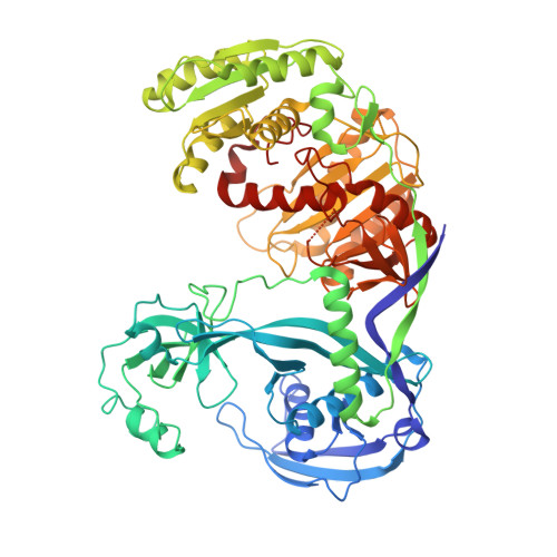
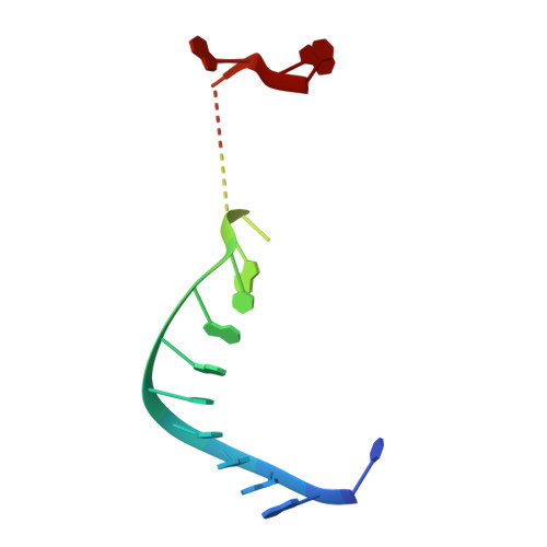
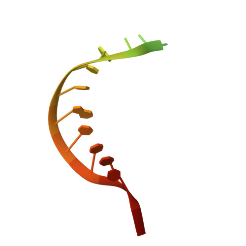
Structure of an argonaute silencing complex with a seed-containing guide DNA and target RNA duplex.

Wang, Y.Juranek, S.Li, H.Sheng, G.Tuschl, T.Patel, D.J.

(2008) Nature 456: 921-926

  • DOI: https://doi.org/10.1038/nature07666
  • Primary Citation of Related Structures:  
    3F73

  • PubMed Abstract: 

    Here we report on a 3.0 A crystal structure of a ternary complex of wild-type Thermus thermophilus argonaute bound to a 5'-phosphorylated 21-nucleotide guide DNA and a 20-nucleotide target RNA containing cleavage-preventing mismatches at the 10-11 step. The seed segment (positions 2 to 8) adopts an A-helical-like Watson-Crick paired duplex, with both ends of the guide strand anchored in the complex. An arginine, inserted between guide-strand bases 10 and 11 in the binary complex, locking it in an inactive conformation, is released on ternary complex formation. The nucleic-acid-binding channel between the PAZ- and PIWI-containing lobes of argonaute widens on formation of a more open ternary complex. The relationship of structure to function was established by determining cleavage activity of ternary complexes containing position-dependent base mismatch, bulge and 2'-O-methyl modifications. Consistent with the geometry of the ternary complex, bulges residing in the seed segments of the target, but not the guide strand, were better accommodated and their complexes were catalytically active.


  • Organizational Affiliation

    Structural Biology Program, Memorial-Sloan Kettering Cancer Center, New York, New York 10065, USA.


Macromolecules

Find similar proteins by:  (by identity cutoff)  |  3D Structure
Entity ID: 1
MoleculeChains Sequence LengthOrganismDetailsImage
ARGONAUTE
A, B
685Thermus thermophilus HB27Mutation(s): 0 
Gene Names: TT_P0026
UniProt
Find proteins for Q746M7 (Thermus thermophilus (strain ATCC BAA-163 / DSM 7039 / HB27))
Explore Q746M7 
Go to UniProtKB:  Q746M7
Entity Groups  
Sequence Clusters30% Identity50% Identity70% Identity90% Identity95% Identity100% Identity
UniProt GroupQ746M7
Sequence Annotations
Expand
  • Reference Sequence

Find similar nucleic acids by:  Sequence   |   3D Structure  

Entity ID: 2
MoleculeChains LengthOrganismImage
DNA (5'-D(P*DTP*DGP*DAP*DGP*DGP*DTP*DAP*DGP*DTP*DAP*DGP*DGP*DTP*DTP*DGP*DTP*DA*DTP*DAP*DGP*DT)-3')C,
E [auth X]
21N/A
Sequence Annotations
Expand
  • Reference Sequence

Find similar nucleic acids by:  Sequence   |   3D Structure  

Entity ID: 3
MoleculeChains LengthOrganismImage
RNA (5'-R(*UP*AP*UP*AP*CP*AP*A*CP*UP*CP*AP*CP*UP*AP*CP*CP*UP*CP*GP*U)-3')D [auth H],
F [auth Y]
20N/A
Sequence Annotations
Expand
  • Reference Sequence
Experimental Data & Validation

Experimental Data

  • Method: X-RAY DIFFRACTION
  • Resolution: 3.00 Å
  • R-Value Free: 0.282 
  • R-Value Work: 0.221 
  • R-Value Observed: 0.221 
  • Space Group: P 1 21 1
Unit Cell:
Length ( Å )Angle ( ˚ )
a = 61.162α = 90
b = 120.527β = 105.3
c = 109.085γ = 90
Software Package:
Software NamePurpose
CNSrefinement
PDB_EXTRACTdata extraction
HKL-2000data reduction
HKL-2000data scaling
PHASESphasing

Structure Validation

View Full Validation Report


Entry History 

Deposition Data

Revision History  (Full details and data files)

  • Version 1.0: 2008-12-16
    Type: Initial release
  • Version 1.1: 2011-07-13
    Changes: Source and taxonomy, Version format compliance
  • Version 1.2: 2023-09-06
    Changes: Data collection, Database references, Derived calculations, Refinement description