3E5C

Crystal Structure of the SMK box (SAM-III) Riboswitch with SAM

  • Classification: RNA
  • Mutation(s): No 

  • Deposited: 2008-08-13 Released: 2008-10-07 
  • Deposition Author(s): Lu, C.

Experimental Data Snapshot

  • Method: X-RAY DIFFRACTION
  • Resolution: 2.25 Å
  • R-Value Free: 0.227 
  • R-Value Work: 0.221 
  • R-Value Observed: 0.221 

wwPDB Validation   3D Report Full Report


Ligand Structure Quality Assessment 


This is version 1.2 of the entry. See complete history


Literature

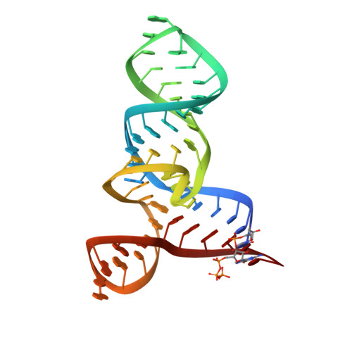
Crystal structures of the SAM-III/S(MK) riboswitch reveal the SAM-dependent translation inhibition mechanism.

Lu, C.Smith, A.M.Fuchs, R.T.Ding, F.Rajashankar, K.Henkin, T.M.Ke, A.

(2008) Nat Struct Mol Biol 15: 1076-1083

  • DOI: https://doi.org/10.1038/nsmb.1494
  • Primary Citation of Related Structures:  
    3E5C, 3E5E, 3E5F

  • PubMed Abstract: 

    Three distinct classes of S-adenosyl-L-methionine (SAM)-responsive riboswitches have been identified that regulate bacterial gene expression at the levels of transcription attenuation or translation inhibition. The S(MK) box (SAM-III) translational riboswitch has been identified in the SAM synthetase gene in members of the Lactobacillales. Here we report the 2.2-A crystal structure of the Enterococcus faecalis S(MK) box riboswitch. The Y-shaped riboswitch organizes its conserved nucleotides around a three-way junction for SAM recognition. The Shine-Dalgarno sequence, which is sequestered by base-pairing with the anti-Shine-Dalgarno sequence in response to SAM binding, also directly participates in SAM recognition. The riboswitch makes extensive interactions with the adenosine and sulfonium moieties of SAM but does not appear to recognize the tail of the methionine moiety. We captured a structural snapshot of the S(MK) box riboswitch sampling the near-cognate ligand S-adenosyl-L-homocysteine (SAH) in which SAH was found to adopt an alternative conformation and fails to make several key interactions.


  • Organizational Affiliation

    Department of Molecular Biology and Genetics, Cornell University, 251 Biotechnology Building, Ithaca, New York 14853, USA.


Macromolecules
Find similar nucleic acids by:  (by identity cutoff)  |  3D Structure
Entity ID: 1
MoleculeChains LengthOrganismImage
SMK box (SAM-III) Riboswitch53N/A
Sequence Annotations
Expand
  • Reference Sequence
Small Molecules
Ligands 2 Unique
IDChains Name / Formula / InChI Key2D Diagram3D Interactions
SAM
Query on SAM

Download Ideal Coordinates CCD File 
Q [auth A]S-ADENOSYLMETHIONINE
C15 H22 N6 O5 S
MEFKEPWMEQBLKI-FCKMPRQPSA-N
SR
Query on SR

Download Ideal Coordinates CCD File 
B [auth A]
C [auth A]
D [auth A]
E [auth A]
F [auth A]
B [auth A],
C [auth A],
D [auth A],
E [auth A],
F [auth A],
G [auth A],
H [auth A],
I [auth A],
J [auth A],
K [auth A],
L [auth A],
M [auth A],
N [auth A],
O [auth A],
P [auth A]
STRONTIUM ION
Sr
PWYYWQHXAPXYMF-UHFFFAOYSA-N
Binding Affinity Annotations 
IDSourceBinding Affinity
SAM PDBBind:  3E5C Kd: 0.57 (nM) from 1 assay(s)
Experimental Data & Validation

Experimental Data

  • Method: X-RAY DIFFRACTION
  • Resolution: 2.25 Å
  • R-Value Free: 0.227 
  • R-Value Work: 0.221 
  • R-Value Observed: 0.221 
  • Space Group: I 41 2 2
Unit Cell:
Length ( Å )Angle ( ˚ )
a = 97.806α = 90
b = 97.806β = 90
c = 87.138γ = 90
Software Package:
Software NamePurpose
CNSrefinement
HKL-2000data collection
HKL-2000data reduction
HKL-2000data scaling
PHASERphasing

Structure Validation

View Full Validation Report


Ligand Structure Quality Assessment 


Entry History 

Deposition Data

  • Released Date: 2008-10-07 
  • Deposition Author(s): Lu, C.

Revision History  (Full details and data files)

  • Version 1.0: 2008-10-07
    Type: Initial release
  • Version 1.1: 2011-07-13
    Changes: Version format compliance
  • Version 1.2: 2024-02-21
    Changes: Data collection, Database references, Derived calculations