3DHS

Mapping metal-binding sites in the catalytic domain of bacterial RNase P RNA


Experimental Data Snapshot

  • Method: X-RAY DIFFRACTION
  • Resolution: 3.60 Å
  • R-Value Free: 0.314 
  • R-Value Work: 0.290 

wwPDB Validation   3D Report Full Report


This is version 1.2 of the entry. See complete history


Literature

Mapping metal-binding sites in the catalytic domain of bacterial RNase P RNA

Kazantsev, A.V.Krivenko, A.A.Pace, N.R.

(2009) RNA 15: 266-276

  • DOI: https://doi.org/10.1261/rna.1331809
  • Primary Citation of Related Structures:  
    3DHS

  • PubMed Abstract: 

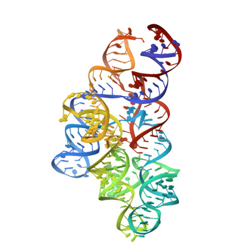
    Ribonuclease P (RNase P) is a ribonucleoprotein enzyme that contains a universally conserved, catalytically active RNA component. RNase P RNA requires divalent metal ions for folding, substrate binding, and catalysis. Despite recent advances in understanding the structure of RNase P RNA, no comprehensive analysis of metal-binding sites has been reported, in part due to the poor crystallization properties of this large RNA. We have developed an abbreviated yet still catalytic construct, Bst P7Delta RNA, which contains the catalytic domain of the bacterial RNase P RNA and has improved crystallization properties. We use this mutant RNA as well as the native RNA to map metal-binding sites in the catalytic core of the bacterial RNase P RNA, by anomalous scattering in diffraction analysis. The results provide insight into the interplay between RNA structure and focalization of metal ions, and a structural basis for some previous biochemical observations with RNase P. We use electrostatic calculations to extract the potential functional significance of these metal-binding sites with respect to binding Mg(2+). The results suggest that with at least one important exception of specific binding, these sites mainly map areas of diffuse association of magnesium ions.


  • Organizational Affiliation

    Department of Molecular, Cellular, and Developmental Biology, University of Colorado at Boulder, 80309, USA.


Macromolecules
Find similar nucleic acids by:  (by identity cutoff)  |  3D Structure
Entity ID: 1
MoleculeChains LengthOrganismImage
RNase P RNA268N/A
Sequence Annotations
Expand
  • Reference Sequence
Small Molecules
Ligands 1 Unique
IDChains Name / Formula / InChI Key2D Diagram3D Interactions
OS
Query on OS

Download Ideal Coordinates CCD File 
B [auth A]
C [auth A]
D [auth A]
E [auth A]
F [auth A]
B [auth A],
C [auth A],
D [auth A],
E [auth A],
F [auth A],
G [auth A],
H [auth A],
I [auth A],
J [auth A],
K [auth A],
L [auth A],
M [auth A],
N [auth A],
O [auth A],
P [auth A]
OSMIUM ION
Os
XQBKHDFIPARBOX-UHFFFAOYSA-N
Experimental Data & Validation

Experimental Data

  • Method: X-RAY DIFFRACTION
  • Resolution: 3.60 Å
  • R-Value Free: 0.314 
  • R-Value Work: 0.290 
  • Space Group: C 2 2 2
Unit Cell:
Length ( Å )Angle ( ˚ )
a = 147.35α = 90
b = 160.01β = 90
c = 133.45γ = 90
Software Package:
Software NamePurpose
d*TREKdata scaling
CNSrefinement
CNSphasing
PDB_EXTRACTdata extraction
ADSCdata collection
d*TREKdata reduction

Structure Validation

View Full Validation Report


Entry History 

Deposition Data

Revision History  (Full details and data files)

  • Version 1.0: 2009-01-27
    Type: Initial release
  • Version 1.1: 2011-07-13
    Changes: Version format compliance
  • Version 1.2: 2023-08-30
    Changes: Data collection, Database references, Derived calculations, Refinement description