3CQL

Crystal Structure of GH family 19 chitinase from Carica papaya


Experimental Data Snapshot

  • Method: X-RAY DIFFRACTION
  • Resolution: 1.50 Å
  • R-Value Free: 0.187 
  • R-Value Work: 0.161 
  • R-Value Observed: 0.162 

wwPDB Validation   3D Report Full Report


Ligand Structure Quality Assessment 


This is version 2.0 of the entry. See complete history


Literature

X-ray Structure of Papaya Chitinase Reveals the Substrate Binding Mode of Glycosyl Hydrolase Family 19 Chitinases.

Huet, J.Rucktooa, P.Clantin, B.Azarkan, M.Looze, Y.Villeret, V.Wintjens, R.

(2008) Biochemistry 47: 8283-8291

  • DOI: https://doi.org/10.1021/bi800655u
  • Primary Citation of Related Structures:  
    3CQL

  • PubMed Abstract: 

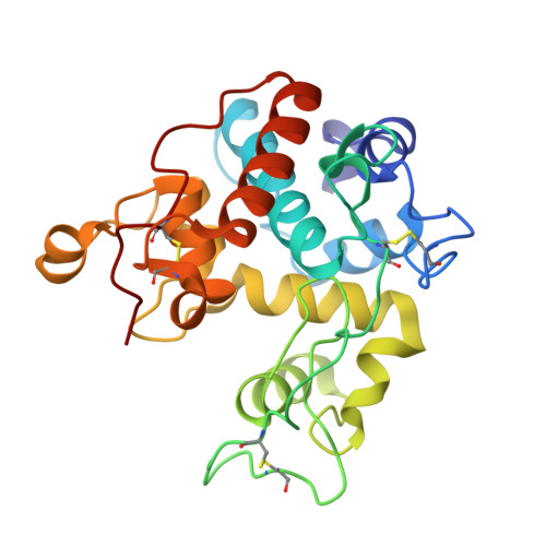
    The crystal structure of a chitinase from Carica papaya has been solved by the molecular replacement method and is reported to a resolution of 1.5 A. This enzyme belongs to family 19 of the glycosyl hydrolases. Crystals have been obtained in the presence of N-acetyl- d-glucosamine (GlcNAc) in the crystallization solution and two well-defined GlcNAc molecules have been identified in the catalytic cleft of the enzyme, at subsites -2 and +1. These GlcNAc moieties bind to the protein via an extensive network of interactions which also involves many hydrogen bonds mediated by water molecules, underlying their role in the catalytic mechanism. A complex of the enzyme with a tetra-GlcNAc molecule has been elaborated, using the experimental interactions observed for the bound GlcNAc saccharides. This model allows to define four major substrate interacting regions in the enzyme, comprising residues located around the catalytic Glu67 (His66 and Thr69), the short segment E89-R90 containing the second catalytic residue Glu89, the region 120-124 (residues Ser120, Trp121, Tyr123, and Asn124), and the alpha-helical segment 198-202 (residues Ile198, Asn199, Gly201, and Leu202). Water molecules from the crystal structure were introduced during the modeling procedure, allowing to pinpoint several additional residues involved in ligand binding that were not previously reported in studies of poly-GlcNAc/family 19 chitinase complexes. This work underlines the role played by water-mediated hydrogen bonding in substrate binding as well as in the catalytic mechanism of the GH family 19 chitinases. Finally, a new sequence motif for family 19 chitinases has been identified between residues Tyr111 and Tyr125.


  • Organizational Affiliation

    Service de Chimie Générale (CP: 206/4), Institut de Pharmacie, Université Libre de Bruxelles (ULB), Campus de la Plaine, Boulevard du Triomphe, B-1050 Brussels, Belgium.


Macromolecules
Find similar proteins by:  (by identity cutoff)  |  3D Structure
Entity ID: 1
MoleculeChains Sequence LengthOrganismDetailsImage
Endochitinase
A, B
243Carica papayaMutation(s): 0 
EC: 3.2.1.14
UniProt
Find proteins for P85084 (Carica papaya)
Explore P85084 
Go to UniProtKB:  P85084
Entity Groups  
Sequence Clusters30% Identity50% Identity70% Identity90% Identity95% Identity100% Identity
UniProt GroupP85084
Sequence Annotations
Expand
  • Reference Sequence
Small Molecules
Ligands 4 Unique
IDChains Name / Formula / InChI Key2D Diagram3D Interactions
NAG
Query on NAG

Download Ideal Coordinates CCD File 
D [auth A],
H [auth B],
I [auth B],
J [auth B]
2-acetamido-2-deoxy-beta-D-glucopyranose
C8 H15 N O6
OVRNDRQMDRJTHS-FMDGEEDCSA-N
NDG
Query on NDG

Download Ideal Coordinates CCD File 
C [auth A],
E [auth A]
2-acetamido-2-deoxy-alpha-D-glucopyranose
C8 H15 N O6
OVRNDRQMDRJTHS-PVFLNQBWSA-N
SO4
Query on SO4

Download Ideal Coordinates CCD File 
F [auth A],
K [auth B],
L [auth B],
M [auth B]
SULFATE ION
O4 S
QAOWNCQODCNURD-UHFFFAOYSA-L
GOL
Query on GOL

Download Ideal Coordinates CCD File 
G [auth A]GLYCEROL
C3 H8 O3
PEDCQBHIVMGVHV-UHFFFAOYSA-N
Binding Affinity Annotations 
IDSourceBinding Affinity
NDG Binding MOAD:  3CQL Ka: 4 (M^-1) from 1 assay(s)
NAG Binding MOAD:  3CQL Ka: 4 (M^-1) from 1 assay(s)
Experimental Data & Validation

Experimental Data

  • Method: X-RAY DIFFRACTION
  • Resolution: 1.50 Å
  • R-Value Free: 0.187 
  • R-Value Work: 0.161 
  • R-Value Observed: 0.162 
  • Space Group: P 1 21 1
Unit Cell:
Length ( Å )Angle ( ˚ )
a = 69α = 90
b = 44.49β = 94.97
c = 76.81γ = 90
Software Package:
Software NamePurpose
MOLREPphasing
REFMACrefinement
PDB_EXTRACTdata extraction
XSCALEdata scaling

Structure Validation

View Full Validation Report


Ligand Structure Quality Assessment 


Entry History 

Deposition Data

Revision History  (Full details and data files)

  • Version 1.0: 2008-08-05
    Type: Initial release
  • Version 1.1: 2011-07-13
    Changes: Non-polymer description, Version format compliance
  • Version 2.0: 2020-07-29
    Type: Remediation
    Reason: Carbohydrate remediation
    Changes: Atomic model, Data collection, Derived calculations, Structure summary