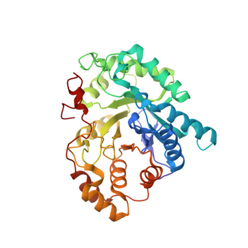
Crystal Structures of Human Delta4-3-Ketosteroid 5beta-Reductase (AKR1D1) Reveal the Presence of an Alternative Binding Site Responsible for Substrate Inhibition (dagger) (,) (double dagger).
Faucher, F., Cantin, L., Luu-The, V., Labrie, F., Breton, R.(2008) Biochemistry 47: 13537-13546
- PubMed: 19075558
- DOI: https://doi.org/10.1021/bi801276h
- Primary Citation of Related Structures:
3CAQ, 3CAS - PubMed Abstract:
The 5beta-reductases (AKR1D1-3) are unique enzymes able to catalyze efficiently and in a stereospecific manner the 5beta-reduction of the C4-C5 double bond found in Delta4-3-ketosteroids, including steroid hormones and bile acids precursors such as 7alpha-hydroxy-4-cholesten-3-one and 7alpha,12alpha-dihydroxy-4-cholesten-3-one. In order to elucidate the binding mode and substrate specificity in detail, biochemical and structural studies on human 5beta-reductase (h5beta-red; AKR1D1) have been recently undertaken. The crystal structure of a h5beta-red binary complex provides a complete picture of the NADPH-enzyme interactions involving the flexible loop B, which contributes to the maintenance of the cofactor in its binding site by acting as a "safety belt". Structural comparison with binary complexes of AKR1C enzymes, specifically the human type 3 3alpha-hydroxysteroid dehydrogenase (AKR1C2) and the mouse 17alpha-hydroxysteroid dehydrogenase (AKR1C21), also revealed particularities in loop B positioning that make the steroid-binding cavity of h5beta-red substantially larger than those of the two other enzymes. Kinetic characterization of the purified recombinant h5beta-red has shown that this enzyme exerts a strong activity toward progesterone (Prog) and androstenedione (Delta4) but is rapidly inhibited by these substrates once their concentrations reach 2-times their K(m) value. A crystal structure of the h5beta-red in ternary complex with NADPH and Delta4 has revealed that the large steroid-binding site of this enzyme also contains a subsite in which the Delta4 molecule is found. When bound in this subsite, Delta4 completely impedes the passage of another substrate molecule toward the catalytic site. The importance of this alternative binding site for the inhibition of h5beta-red was finally proven by site-directed mutagenesis, which demonstrated that the replacement of one of the residues delineating this site (Val(309)) by a phenylalanine completely abolishes the substrate inhibition. The results of this report provide structural insights into the substrate inhibition of h5beta-red by C19- and C21-steroids.
Organizational Affiliation:
Oncology and Molecular Endocrinology Research Center, Laval University Medical Center (CHUL) and Laval University, Laval, Quebec (QC) G1V 4G2, Canada.