3BTA

CRYSTAL STRUCTURE OF BOTULINUM NEUROTOXIN SEROTYPE A


Experimental Data Snapshot

  • Method: X-RAY DIFFRACTION
  • Resolution: 3.20 Å
  • R-Value Free: 0.280 
  • R-Value Work: 0.220 
  • R-Value Observed: 0.220 

wwPDB Validation   3D Report Full Report


This is version 1.3 of the entry. See complete history


Literature

Crystal structure of botulinum neurotoxin type A and implications for toxicity.

Lacy, D.B.Tepp, W.Cohen, A.C.DasGupta, B.R.Stevens, R.C.

(1998) Nat Struct Biol 5: 898-902

  • DOI: https://doi.org/10.1038/2338
  • Primary Citation of Related Structures:  
    3BTA

  • PubMed Abstract: 

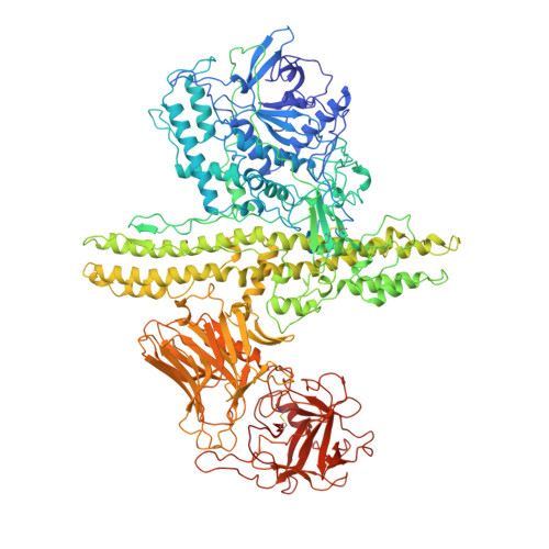
    Botulinum neurotoxin type A (BoNT/A) is the potent disease agent in botulism, a potential biological weapon and an effective therapeutic drug for involuntary muscle disorders. The crystal structure of the entire 1,285 amino acid di-chain neurotoxin was determined at 3.3 A resolution. The structure reveals that the translocation domain contains a central pair of alpha-helices 105 A long and a approximately 50 residue loop or belt that wraps around the catalytic domain. This belt partially occludes a large channel leading to a buried, negative active site--a feature that calls for radically different inhibitor design strategies from those currently used. The fold of the translocation domain suggests a mechanism of pore formation different from other toxins. Lastly, the toxin appears as a hybrid of varied structural motifs and suggests a modular assembly of functional subunits to yield pathogenesis.


  • Organizational Affiliation

    Department of Chemistry and Earnest Orlando Lawrence Berkeley National Laboratory, University of California, Berkeley, 94720, USA.


Macromolecules
Find similar proteins by:  (by identity cutoff)  |  3D Structure
Entity ID: 1
MoleculeChains Sequence LengthOrganismDetailsImage
PROTEIN (BOTULINUM NEUROTOXIN TYPE A)1,295Clostridium botulinumMutation(s): 0 
EC: 3.4.24.69
UniProt
Find proteins for P0DPI1 (Clostridium botulinum (strain Hall / ATCC 3502 / NCTC 13319 / Type A))
Explore P0DPI1 
Go to UniProtKB:  P0DPI1
Entity Groups  
Sequence Clusters30% Identity50% Identity70% Identity90% Identity95% Identity100% Identity
UniProt GroupP0DPI1
Sequence Annotations
Expand
  • Reference Sequence
Small Molecules
Ligands 1 Unique
IDChains Name / Formula / InChI Key2D Diagram3D Interactions
ZN
Query on ZN

Download Ideal Coordinates CCD File 
B [auth A]ZINC ION
Zn
PTFCDOFLOPIGGS-UHFFFAOYSA-N
Experimental Data & Validation

Experimental Data

  • Method: X-RAY DIFFRACTION
  • Resolution: 3.20 Å
  • R-Value Free: 0.280 
  • R-Value Work: 0.220 
  • R-Value Observed: 0.220 
  • Space Group: P 31 2 1
Unit Cell:
Length ( Å )Angle ( ˚ )
a = 170.12α = 90
b = 170.12β = 90
c = 161γ = 120
Software Package:
Software NamePurpose
DENZOdata reduction
SCALEPACKdata scaling
MLPHAREphasing
CNSrefinement

Structure Validation

View Full Validation Report


Entry History 

Deposition Data

Revision History  (Full details and data files)

  • Version 1.0: 1999-10-01
    Type: Initial release
  • Version 1.1: 2008-04-27
    Changes: Version format compliance
  • Version 1.2: 2011-07-13
    Changes: Version format compliance
  • Version 1.3: 2023-12-27
    Changes: Data collection, Database references, Derived calculations