3B31

Crystal structure of domain III of the Cricket Paralysis Virus IRES RNA


Experimental Data Snapshot

  • Method: X-RAY DIFFRACTION
  • Resolution: 2.40 Å
  • R-Value Free: 0.269 
  • R-Value Work: 0.239 

wwPDB Validation   3D Report Full Report


This is version 1.3 of the entry. See complete history


Literature

tRNA-mRNA mimicry drives translation initiation from a viral IRES.

Costantino, D.A.Pfingsten, J.S.Rambo, R.P.Kieft, J.S.

(2008) Nat Struct Mol Biol 15: 57-64

  • DOI: https://doi.org/10.1038/nsmb1351
  • Primary Citation of Related Structures:  
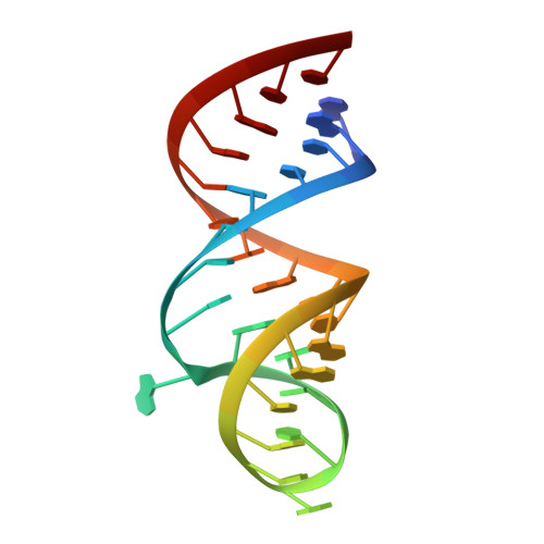
    3B31

  • PubMed Abstract: 

    Internal ribosome entry site (IRES) RNAs initiate protein synthesis in eukaryotic cells by a noncanonical cap-independent mechanism. IRESes are critical for many pathogenic viruses, but efforts to understand their function are complicated by the diversity of IRES sequences as well as by limited high-resolution structural information. The intergenic region (IGR) IRESes of the Dicistroviridae viruses are powerful model systems to begin to understand IRES function. Here we present the crystal structure of a Dicistroviridae IGR IRES domain that interacts with the ribosome's decoding groove. We find that this RNA domain precisely mimics the transfer RNA anticodon-messenger RNA codon interaction, and its modeled orientation on the ribosome helps explain translocation without peptide bond formation. When combined with a previous structure, this work completes the first high-resolution description of an IRES RNA and provides insight into how RNAs can manipulate complex biological machines.


  • Organizational Affiliation

    Department of Biochemistry and Molecular Genetics, University of Colorado at Denver and Health Sciences Center, Mail Stop 8101, PO Box 6511, Aurora, Colorado 80045, USA.


Macromolecules
Find similar nucleic acids by:  (by identity cutoff)  |  3D Structure
Entity ID: 1
MoleculeChains LengthOrganismImage
RNA (29-MER)29N/A
Sequence Annotations
Expand
  • Reference Sequence

Find similar nucleic acids by:  Sequence   |   3D Structure  

Entity ID: 2
MoleculeChains LengthOrganismImage
RNA (5'-R(P*UP*AP*AP*GP*AP*AP*AP*UP*UP*UP*AP*CP*CP*U)-3')14N/A
Sequence Annotations
Expand
  • Reference Sequence
Experimental Data & Validation

Experimental Data

  • Method: X-RAY DIFFRACTION
  • Resolution: 2.40 Å
  • R-Value Free: 0.269 
  • R-Value Work: 0.239 
  • Space Group: P 41 21 2
Unit Cell:
Length ( Å )Angle ( ˚ )
a = 58.724α = 90
b = 58.724β = 90
c = 98.989γ = 90
Software Package:
Software NamePurpose
Blu-Icedata collection
PHENIXmodel building
CNSrefinement
ELVESdata reduction
MOSFLMdata reduction
CCP4data scaling
PHENIXphasing

Structure Validation

View Full Validation Report


Entry History 

Deposition Data

Revision History  (Full details and data files)

  • Version 1.0: 2007-12-25
    Type: Initial release
  • Version 1.1: 2011-07-13
    Changes: Version format compliance
  • Version 1.2: 2017-10-25
    Changes: Refinement description
  • Version 1.3: 2024-02-21
    Changes: Data collection, Database references, Derived calculations