3AVY

Structure of viral RNA polymerase complex 6


Experimental Data Snapshot

  • Method: X-RAY DIFFRACTION
  • Resolution: 2.62 Å
  • R-Value Free: 0.262 
  • R-Value Work: 0.214 
  • R-Value Observed: 0.216 

wwPDB Validation   3D Report Full Report


Ligand Structure Quality Assessment 


This is version 1.3 of the entry. See complete history


Literature

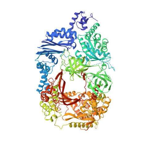
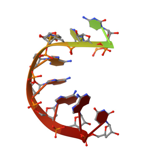
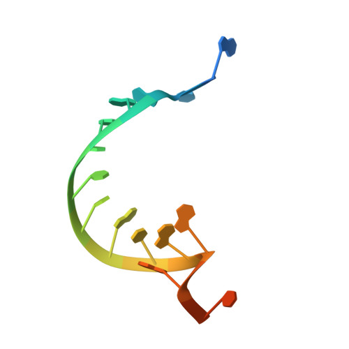
Molecular basis for RNA polymerization by Q beta replicase

Takeshita, D.Tomita, K.

(2012) Nat Struct Mol Biol 19: 229-237

  • DOI: https://doi.org/10.1038/nsmb.2204
  • Primary Citation of Related Structures:  
    3AVT, 3AVU, 3AVV, 3AVW, 3AVX, 3AVY

  • PubMed Abstract: 

    Core Qβ replicase comprises the Qβ virus-encoded RNA-dependent RNA polymerase (β-subunit) and the host Escherichia coli translational elongation factors EF-Tu and EF-Ts. The functions of the host proteins in the viral replicase are not clear. Structural analyses of RNA polymerization by core Qβ replicase reveal that at the initiation stage, the 3'-adenine of the template RNA provides a stable platform for de novo initiation. EF-Tu in Qβ replicase forms a template exit channel with the β-subunit. At the elongation stages, the C-terminal region of the β-subunit, assisted by EF-Tu, splits the temporarily double-stranded RNA between the template and nascent RNAs before translocation of the single-stranded template RNA into the exit channel. Therefore, EF-Tu in Qβ replicase modulates RNA elongation processes in a distinct manner from its established function in protein synthesis.


  • Organizational Affiliation

    Biomedical Research Institute, National Institute of Advanced Industrial Science and Technology, Ibaraki, Japan.


Macromolecules

Find similar proteins by:  (by identity cutoff)  |  3D Structure
Entity ID: 1
MoleculeChains Sequence LengthOrganismDetailsImage
Elongation factor Ts, Elongation factor Tu, LINKER, Q beta replicase1,289Escherichia coli O157:H7synthetic constructQubevirus durumMutation(s): 0 
UniProt
Find proteins for P14647 (Escherichia virus Qbeta)
Explore P14647 
Go to UniProtKB:  P14647
Find proteins for P0A6N3 (Escherichia coli O157:H7)
Explore P0A6N3 
Go to UniProtKB:  P0A6N3
Find proteins for P0A6P3 (Escherichia coli O157:H7)
Explore P0A6P3 
Go to UniProtKB:  P0A6P3
Entity Groups  
Sequence Clusters30% Identity50% Identity70% Identity90% Identity95% Identity100% Identity
UniProt GroupsP14647P0A6P3P0A6N3
Sequence Annotations
Expand
  • Reference Sequence

Find similar nucleic acids by:  Sequence   |   3D Structure  

Entity ID: 2
MoleculeChains LengthOrganismImage
RNA (5'-R(*GP*GP*GP*UP*CP*CP*AP*UP*AP*AP*AP*AP*U)-3')B [auth G]13N/A
Sequence Annotations
Expand
  • Reference Sequence

Find similar nucleic acids by:  Sequence   |   3D Structure  

Entity ID: 3
MoleculeChains LengthOrganismImage
RNA (5'-R(*AP*AP*CP*GP*AP*UP*UP*UP*UP*AP*UP*GP*GP*AP*CP*CP*CP*A)-3')C [auth T]18N/A
Sequence Annotations
Expand
  • Reference Sequence
Experimental Data & Validation

Experimental Data

  • Method: X-RAY DIFFRACTION
  • Resolution: 2.62 Å
  • R-Value Free: 0.262 
  • R-Value Work: 0.214 
  • R-Value Observed: 0.216 
  • Space Group: C 2 2 21
Unit Cell:
Length ( Å )Angle ( ˚ )
a = 140.07α = 90
b = 257.22β = 90
c = 101.31γ = 90
Software Package:
Software NamePurpose
HKL-2000data collection
MOLREPphasing
PHENIXrefinement
HKL-2000data reduction
HKL-2000data scaling

Structure Validation

View Full Validation Report


Ligand Structure Quality Assessment 


Entry History 

Deposition Data

Revision History  (Full details and data files)

  • Version 1.0: 2012-01-18
    Type: Initial release
  • Version 1.1: 2013-09-11
    Changes: Database references
  • Version 1.2: 2017-06-28
    Changes: Source and taxonomy
  • Version 1.3: 2023-11-01
    Changes: Data collection, Database references, Derived calculations, Refinement description