2ZHH

Crystal structure of SoxR


Experimental Data Snapshot

  • Method: X-RAY DIFFRACTION
  • Resolution: 3.20 Å
  • R-Value Free: 0.276 
  • R-Value Work: 0.244 
  • R-Value Observed: 0.244 

wwPDB Validation   3D Report Full Report


This is version 1.2 of the entry. See complete history


Literature

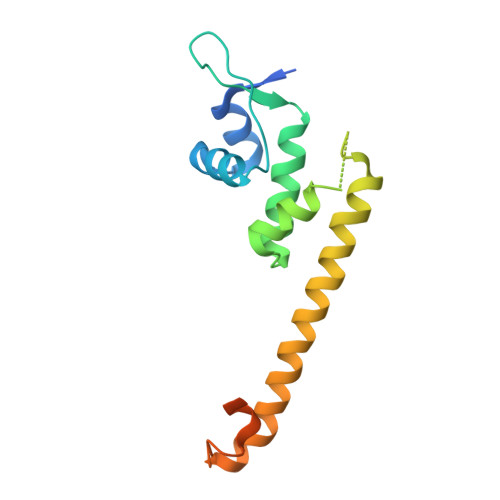
Crystal structure of the [2Fe-2S] oxidative-stress sensor SoxR bound to DNA

Watanabe, S.Kita, A.Kobayashi, K.Miki, K.

(2008) Proc Natl Acad Sci U S A 105: 4121-4126

  • DOI: https://doi.org/10.1073/pnas.0709188105
  • Primary Citation of Related Structures:  
    2ZHG, 2ZHH

  • PubMed Abstract: 

    The [2Fe-2S] transcription factor SoxR, a member of the MerR family, functions as a bacterial sensor of oxidative stress such as superoxide and nitric oxide. SoxR is activated by reversible one-electron oxidation of the [2Fe-2S] cluster and then enhances the production of various antioxidant proteins through the soxRS regulon. In the active state, SoxR and other MerR family proteins activate transcription from unique promoters, which have a long 19- or 20-bp spacer between the -35 and -10 operator elements, by untwisting the promoter DNA. Here, we show the crystal structures of SoxR and its complex with the target promoter in the oxidized (active) state. The structures reveal that the [2Fe-2S] cluster of SoxR is completely solvent-exposed and surrounded by an asymmetric environment stabilized by interaction with the other subunit. The asymmetrically charged environment of the [2Fe-2S] cluster probably causes redox-dependent conformational changes of SoxR and the target promoter. Compared with the promoter structures with the 19-bp spacer previously studied, the DNA structure is more sharply bent, by approximately 1 bp, with the two central base pairs holding Watson-Crick base pairs. Comparison of the target promoter sequences of the MerR family indicates that the present DNA structure represents the activated conformation of the target promoter with a 20-bp spacer in the MerR family.


  • Organizational Affiliation

    Department of Chemistry, Graduate School of Science, Kyoto University, Sakyo-ku, Kyoto 606-8502, Japan.


Macromolecules
Find similar proteins by:  (by identity cutoff)  |  3D Structure
Entity ID: 1
MoleculeChains Sequence LengthOrganismDetailsImage
Redox-sensitive transcriptional activator soxR154Escherichia coli K-12Mutation(s): 0 
Gene Names: soxR
UniProt
Find proteins for P0ACS2 (Escherichia coli (strain K12))
Explore P0ACS2 
Go to UniProtKB:  P0ACS2
Entity Groups  
Sequence Clusters30% Identity50% Identity70% Identity90% Identity95% Identity100% Identity
UniProt GroupP0ACS2
Sequence Annotations
Expand
  • Reference Sequence
Small Molecules
Ligands 2 Unique
IDChains Name / Formula / InChI Key2D Diagram3D Interactions
FES
Query on FES

Download Ideal Coordinates CCD File 
B [auth A]FE2/S2 (INORGANIC) CLUSTER
Fe2 S2
NIXDOXVAJZFRNF-UHFFFAOYSA-N
DTT
Query on DTT

Download Ideal Coordinates CCD File 
C [auth A]2,3-DIHYDROXY-1,4-DITHIOBUTANE
C4 H10 O2 S2
VHJLVAABSRFDPM-IMJSIDKUSA-N
Experimental Data & Validation

Experimental Data

  • Method: X-RAY DIFFRACTION
  • Resolution: 3.20 Å
  • R-Value Free: 0.276 
  • R-Value Work: 0.244 
  • R-Value Observed: 0.244 
  • Space Group: P 62
Unit Cell:
Length ( Å )Angle ( ˚ )
a = 80.006α = 90
b = 80.006β = 90
c = 88.118γ = 120
Software Package:
Software NamePurpose
CNSrefinement
HKL-2000data collection
HKL-2000data reduction
HKL-2000data scaling
MOLREPphasing

Structure Validation

View Full Validation Report


Entry History 

Deposition Data

Revision History  (Full details and data files)

  • Version 1.0: 2008-03-25
    Type: Initial release
  • Version 1.1: 2011-07-13
    Changes: Source and taxonomy, Version format compliance
  • Version 1.2: 2023-11-01
    Changes: Data collection, Database references, Derived calculations, Refinement description