2XNR

Structural insights into cis element recognition of non- polyadenylated RNAs by the Nab3-RRM


Experimental Data Snapshot

  • Method: X-RAY DIFFRACTION
  • Resolution: 1.60 Å
  • R-Value Free: 0.184 
  • R-Value Work: 0.160 
  • R-Value Observed: 0.162 

wwPDB Validation   3D Report Full Report


This is version 1.3 of the entry. See complete history


Literature

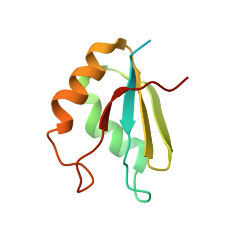
Structural Insights Into Cis Element Recognition of Non-Polyadenylated Rnas by the Nab3-Rrm.

Lunde, B.M.Horner, M.Meinhart, A.

(2011) Nucleic Acids Res 39: 337

  • DOI: https://doi.org/10.1093/nar/gkq751
  • Primary Citation of Related Structures:  
    2XNQ, 2XNR

  • PubMed Abstract: 

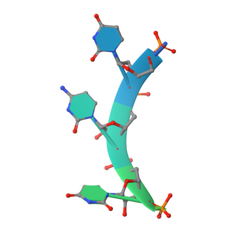
    Transcription termination of non-polyadenylated RNAs in Saccharomyces cerevisiae occurs through the action of the Nrd1-Nab3-Sen1 complex. Part of the decision to terminate via this pathway occurs via direct recognition of sequences within the nascent transcript by RNA recognition motifs (RRMs) within Nrd1 and Nab3. Here we present the 1.6 Å structure of Nab3-RRM bound to its UCUU recognition sequence. The crystal structure reveals clear density for a UCU trinucleotide and a fourth putative U binding site. Nab3-RRM establishes a clear preference for the central cytidine of the UCUU motif, which forms pseudo-base pairing interactions primarily through hydrogen bonds to main chain atoms and one serine hydroxyl group. Specificity for the flanking uridines is less defined; however, binding experiments confirm that these residues are also important for high affinity binding. Comparison of the Nab3-RRM to other structures of RRMs bound to polypyrimidine RNAs showed that this mode of recognition is similar to what is observed for the polypyrimidine-tract binding RRMs, and that the serine residue involved in pseudo-base pairing is only found in RRMs that bind to polypyrimidine RNAs that contain a cytosine base, suggesting a possible mechanism for discriminating between cytosine and uracil bases in RRMs that bind to polypyrimidine-containing RNA.


  • Organizational Affiliation

    Department of Biomolecular Mechanisms, Max Planck Institute for Medical Research, Heidelberg, Germany.


Macromolecules

Find similar proteins by:  (by identity cutoff)  |  3D Structure
Entity ID: 1
MoleculeChains Sequence LengthOrganismDetailsImage
NUCLEAR POLYADENYLATED RNA-BINDING PROTEIN 397Saccharomyces cerevisiaeMutation(s): 0 
UniProt
Find proteins for P38996 (Saccharomyces cerevisiae (strain ATCC 204508 / S288c))
Explore P38996 
Go to UniProtKB:  P38996
Entity Groups  
Sequence Clusters30% Identity50% Identity70% Identity90% Identity95% Identity100% Identity
UniProt GroupP38996
Sequence Annotations
Expand
  • Reference Sequence

Find similar nucleic acids by:  Sequence   |   3D Structure  

Entity ID: 2
MoleculeChains LengthOrganismImage
5'-R(*UP*UP*CP*UP*UP*AP*UP*UP*CP*UP*UP*A)-3'B [auth C]12Saccharomyces cerevisiae
Sequence Annotations
Expand
  • Reference Sequence
Experimental Data & Validation

Experimental Data

  • Method: X-RAY DIFFRACTION
  • Resolution: 1.60 Å
  • R-Value Free: 0.184 
  • R-Value Work: 0.160 
  • R-Value Observed: 0.162 
  • Space Group: P 41
Unit Cell:
Length ( Å )Angle ( ˚ )
a = 30.64α = 90
b = 30.64β = 90
c = 82.42γ = 90
Software Package:
Software NamePurpose
REFMACrefinement
XDSdata reduction
XDSdata scaling
PHASERphasing

Structure Validation

View Full Validation Report


Entry History 

Deposition Data

Revision History  (Full details and data files)

  • Version 1.0: 2010-09-08
    Type: Initial release
  • Version 1.1: 2011-05-08
    Changes: Version format compliance
  • Version 1.2: 2011-07-13
    Changes: Version format compliance
  • Version 1.3: 2023-12-20
    Changes: Data collection, Database references, Derived calculations, Other, Refinement description