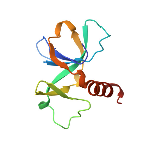
2VUB

CCDB, A TOPOISOMERASE POISON FROM E. COLI


Experimental Data Snapshot

  • Method: X-RAY DIFFRACTION
  • Resolution: 2.45 Å
  • R-Value Free: 0.250 
  • R-Value Work: 0.205 
  • R-Value Observed: 0.205 

wwPDB Validation   3D Report Full Report


This is version 1.4 of the entry. See complete history


Literature

Crystal structure of CcdB, a topoisomerase poison from E. coli.

Loris, R.Dao-Thi, M.-H.Bahassi, E.M.Van Melderen, L.Poortmans, F.Liddington, R.Couturier, M.Wyns, L.

(1999) J Mol Biol 285: 1667-1677

  • DOI: https://doi.org/10.1006/jmbi.1998.2395
  • Primary Citation of Related Structures:  
    1VUB, 2VUB, 3VUB, 4VUB

  • PubMed Abstract: 

    The crystal structure of CcdB, a protein that poisons Escherichia coli gyrase, was determined in three crystal forms. The protein consists of a five-stranded antiparallel beta-pleated sheet followed by a C-terminal alpha-helix. In one of the loops of the sheet, a second small three-stranded antiparallel beta-sheet is inserted that sticks out of the molecule as a wing. This wing contains the LysC proteolytic cleavage site that is protected by CcdA and, therefore, forms a likely CcdA recognition site. A dimer is formed by sheet extension and by extensive hydrophobic contacts involving three of the five methionine residues and the C terminus of the alpha-helix. The surface of the dimer on the side of the alpha-helix is overall negatively charged, while the opposite side as well as the wing sheet is dominated by positive charges. We propose that the CcdB dimer binds into the central hole of the 59 kDa N-terminal fragment of GyrA, after disruption of the head dimer interface of GyrA.


  • Organizational Affiliation

    Laboratorium voor Ultrastructuur, Vlaams Interuniversitair Instituut voor Biotechnologie, Vrije Universiteit Brussel, Paardenstraat 65, Sint-Genesius-Rode, B-1640, Belgium. reloris@vub.ac.be


Macromolecules
Find similar proteins by:  (by identity cutoff)  |  3D Structure
Entity ID: 1
MoleculeChains Sequence LengthOrganismDetailsImage
CCDB
A, B, C, D, E
A, B, C, D, E, F, G, H
101Escherichia coliMutation(s): 0 
Gene Names: CCDB
UniProt
Find proteins for P62554 (Escherichia coli (strain K12))
Explore P62554 
Go to UniProtKB:  P62554
Entity Groups  
Sequence Clusters30% Identity50% Identity70% Identity90% Identity95% Identity100% Identity
UniProt GroupP62554
Sequence Annotations
Expand
  • Reference Sequence
Experimental Data & Validation

Experimental Data

  • Method: X-RAY DIFFRACTION
  • Resolution: 2.45 Å
  • R-Value Free: 0.250 
  • R-Value Work: 0.205 
  • R-Value Observed: 0.205 
  • Space Group: P 21 21 21
Unit Cell:
Length ( Å )Angle ( ˚ )
a = 77.71α = 90
b = 92.98β = 90
c = 142.05γ = 90
Software Package:
Software NamePurpose
X-PLORmodel building
X-PLORrefinement
DENZOdata reduction
SCALEPACKdata scaling
X-PLORphasing

Structure Validation

View Full Validation Report


Entry History 

Deposition Data

Revision History  (Full details and data files)

  • Version 1.0: 1998-06-17
    Type: Initial release
  • Version 1.1: 2008-03-25
    Changes: Version format compliance
  • Version 1.2: 2011-07-13
    Changes: Version format compliance
  • Version 1.3: 2024-02-21
    Changes: Data collection, Database references, Derived calculations, Other
  • Version 1.4: 2024-04-03
    Changes: Refinement description