2QWY

SAM-II riboswitch bound to S-adenosylmethionine


Experimental Data Snapshot

  • Method: X-RAY DIFFRACTION
  • Resolution: 2.80 Å
  • R-Value Free: 0.263 
  • R-Value Work: 0.206 
  • R-Value Observed: 0.206 

wwPDB Validation   3D Report Full Report


Ligand Structure Quality Assessment 


This is version 1.2 of the entry. See complete history


Literature

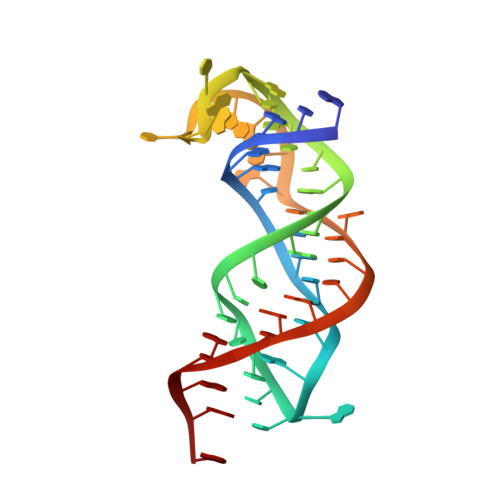
Structure of the SAM-II riboswitch bound to S-adenosylmethionine.

Gilbert, S.D.Rambo, R.P.Van Tyne, D.Batey, R.T.

(2008) Nat Struct Mol Biol 15: 177-182

  • DOI: https://doi.org/10.1038/nsmb.1371
  • Primary Citation of Related Structures:  
    2QWY

  • PubMed Abstract: 

    In bacteria, numerous genes harbor regulatory elements in the 5' untranslated regions of their mRNA, termed riboswitches, which control gene expression by binding small-molecule metabolites. These sequences influence the secondary and tertiary structure of the RNA in a ligand-dependent manner, thereby directing its transcription or translation. The crystal structure of an S-adenosylmethionine-responsive riboswitch found predominantly in proteobacteria, SAM-II, has been solved to reveal a second means by which RNA interacts with this important cellular metabolite. Notably, this is the first structure of a complete riboswitch containing all sequences associated with both the ligand binding aptamer domain and the regulatory expression platform. Chemical probing of this RNA in the absence and presence of ligand shows how the structure changes in response to S-adenosylmethionine to sequester the ribosomal binding site and affect translational gene regulation.


  • Organizational Affiliation

    Department of Chemistry and Biochemistry, Campus Box 215, University of Colorado, Boulder, Colorado 80309, USA.


Macromolecules
Find similar nucleic acids by:  (by identity cutoff)  |  3D Structure
Entity ID: 1
MoleculeChains LengthOrganismImage
SAM-II riboswitch
A, B, C
52N/A
Sequence Annotations
Expand
  • Reference Sequence
Small Molecules
Ligands 4 Unique
IDChains Name / Formula / InChI Key2D Diagram3D Interactions
SAM
Query on SAM

Download Ideal Coordinates CCD File 
H [auth A],
K [auth B],
O [auth C]
S-ADENOSYLMETHIONINE
C15 H22 N6 O5 S
MEFKEPWMEQBLKI-FCKMPRQPSA-N
CS
Query on CS

Download Ideal Coordinates CCD File 
F [auth A],
N [auth C]
CESIUM ION
Cs
NCMHKCKGHRPLCM-UHFFFAOYSA-N
PO4
Query on PO4

Download Ideal Coordinates CCD File 
D [auth A]
E [auth A]
I [auth B]
J [auth B]
L [auth C]
D [auth A],
E [auth A],
I [auth B],
J [auth B],
L [auth C],
M [auth C]
PHOSPHATE ION
O4 P
NBIIXXVUZAFLBC-UHFFFAOYSA-K
MG
Query on MG

Download Ideal Coordinates CCD File 
G [auth A]MAGNESIUM ION
Mg
JLVVSXFLKOJNIY-UHFFFAOYSA-N
Experimental Data & Validation

Experimental Data

  • Method: X-RAY DIFFRACTION
  • Resolution: 2.80 Å
  • R-Value Free: 0.263 
  • R-Value Work: 0.206 
  • R-Value Observed: 0.206 
  • Space Group: C 1 2 1
Unit Cell:
Length ( Å )Angle ( ˚ )
a = 115.258α = 90
b = 48.095β = 108.25
c = 109.617γ = 90
Software Package:
Software NamePurpose
CNSrefinement
CrystalCleardata collection
HKL-2000data reduction
HKL-2000data scaling
SOLVEphasing

Structure Validation

View Full Validation Report


Ligand Structure Quality Assessment 


Entry History 

Deposition Data

Revision History  (Full details and data files)

  • Version 1.0: 2008-01-22
    Type: Initial release
  • Version 1.1: 2011-07-13
    Changes: Version format compliance
  • Version 1.2: 2024-02-21
    Changes: Data collection, Database references, Derived calculations