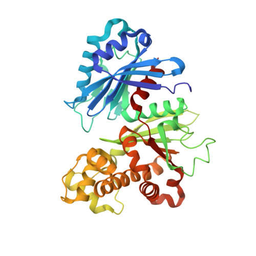
The Crystal Structure of Trypanosoma cruzi Glucokinase Reveals Features Determining Oligomerization and Anomer Specificity of Hexose-phosphorylating Enzymes.
Cordeiro, A.T., Caceres, A.J., Vertommen, D., Concepcion, J.L., Michels, P.A., Versees, W.(2007) J Mol Biol 372: 1215-1226
- PubMed: 17761195
- DOI: https://doi.org/10.1016/j.jmb.2007.07.021
- Primary Citation of Related Structures:
2Q2R - PubMed Abstract:
Glucose is an essential substrate for Trypanosoma cruzi, the protozoan organism responsible for Chagas' disease. The glucose is intracellularly phosphorylated to glucose 6-phosphate. Previously, a hexokinase responsible for this phosphorylation has been characterized. Recently, we identified an ATP-dependent glucokinase in T. cruzi exhibiting a tenfold lower substrate affinity compared to the hexokinase. Both enzymes, which belong to very different groups of the same family, are located inside glycosomes, the peroxisome-like organelles of Kinetoplastida that are known to contain the first seven glycolytic steps as well as enzymes of the oxidative branch of the pentose phosphate pathway. Here, we present the crystallographic structure of T. cruzi glucokinase, in complex with glucose and ADP. The structure suggests a loose tetrameric assembly formed by the association of two tight dimers. TcGlcK was previously reported to exist in a concentration-dependent equilibrium of monomeric and dimeric states. Here, we used mass spectrometry analysis to confirm the existence of TcGlcK monomeric and dimeric states. The analysis of subunit interactions and comparison with the bacterial glucokinases give insights into the forces promoting the stability of the different oligomeric states. Each T. cruzi glucokinase monomer contains one glucose and one ADP molecule. In contrast to hexokinases, which show a moderate preference for the alpha anomer of glucose, the electron density clearly shows the d-glucose bound in the beta configuration in the T.cruzi glucokinase. Kinetic assays with alpha and beta-d-glucose further confirm a moderate preference of the T. cruzi glucokinase for the beta anomer. Structural comparison of the glucokinase and hexokinases permits the identification of a possible mechanism for anomer selectivity in these hexose-phosphorylating enzymes. The preference for distinct anomers suggests that in T. cruzi hexokinase and glucokinase are not directly competing for the same substrate and are probably both present because they exert distinct physiological functions.
Organizational Affiliation:
Research Unit for Tropical Diseases, Christian de Duve Institute of Cellular Pathology, Avenue Hippocrate 74, B-1200 Brussels, Belgium.