2ONJ

Structure of the multidrug ABC transporter Sav1866 from S. aureus in complex with AMP-PNP


Experimental Data Snapshot

  • Method: X-RAY DIFFRACTION
  • Resolution: 3.40 Å
  • R-Value Free: 0.278 
  • R-Value Work: 0.254 

wwPDB Validation   3D Report Full Report


Ligand Structure Quality Assessment 


This is version 1.3 of the entry. See complete history


Literature

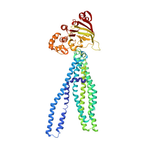
Structure of the multidrug ABC transporter Sav1866 from Staphylococcus aureus in complex with AMP-PNP.

Dawson, R.J.P.Locher, K.P.

(2007) FEBS Lett 581: 935-938

  • DOI: https://doi.org/10.1016/j.febslet.2007.01.073
  • Primary Citation of Related Structures:  
    2ONJ

  • PubMed Abstract: 

    Staphylococcus aureus Sav1866 is a bacterial homolog of the human ABC transporter Mdr1 that causes multidrug resistance in cancer cells. We report the crystal structure of Sav1866 in complex with adenosine-5'-(beta,gamma-imido)triphosphate (AMP-PNP) at 3.4A resolution and compare it with the previously determined structure of Sav1866 with bound ADP. Besides differences in the ATP-binding sites, no significant conformational changes were observed. The results confirm that the ATP-bound state of multidrug ABC transporters is coupled to an outward-facing conformation of the transmembrane domains.


  • Organizational Affiliation

    Institute of Molecular Biology and Biophysics, ETH Zurich HPK D14.3, 8093 Zurich, Switzerland.


Macromolecules
Find similar proteins by:  (by identity cutoff)  |  3D Structure
Entity ID: 1
MoleculeChains Sequence LengthOrganismDetailsImage
Multidrug export ATP-binding/permease protein SAV1866
A, B
578Staphylococcus aureusMutation(s): 0 
EC: 3.6.3
Membrane Entity: Yes 
UniProt
Find proteins for Q99T13 (Staphylococcus aureus (strain Mu50 / ATCC 700699))
Explore Q99T13 
Go to UniProtKB:  Q99T13
Entity Groups  
Sequence Clusters30% Identity50% Identity70% Identity90% Identity95% Identity100% Identity
UniProt GroupQ99T13
Sequence Annotations
Expand
  • Reference Sequence
Experimental Data & Validation

Experimental Data

  • Method: X-RAY DIFFRACTION
  • Resolution: 3.40 Å
  • R-Value Free: 0.278 
  • R-Value Work: 0.254 
  • Space Group: C 1 2 1
Unit Cell:
Length ( Å )Angle ( ˚ )
a = 160.961α = 90
b = 104.447β = 98.23
c = 181.391γ = 90
Software Package:
Software NamePurpose
CNSrefinement
PDB_EXTRACTdata extraction

Structure Validation

View Full Validation Report


Ligand Structure Quality Assessment 


Entry History 

Deposition Data

Revision History  (Full details and data files)

  • Version 1.0: 2007-03-13
    Type: Initial release
  • Version 1.1: 2008-05-01
    Changes: Version format compliance
  • Version 1.2: 2011-07-13
    Changes: Version format compliance
  • Version 1.3: 2023-12-27
    Changes: Data collection, Database references, Derived calculations