2OIY

Crystal structure of the duplex form of the HIV-1(LAI) RNA dimerization initiation site


Experimental Data Snapshot

  • Method: X-RAY DIFFRACTION
  • Resolution: 1.60 Å
  • R-Value Free: 0.250 
  • R-Value Work: 0.242 
  • R-Value Observed: 0.242 

wwPDB Validation   3D Report Full Report


This is version 1.3 of the entry. See complete history


Literature

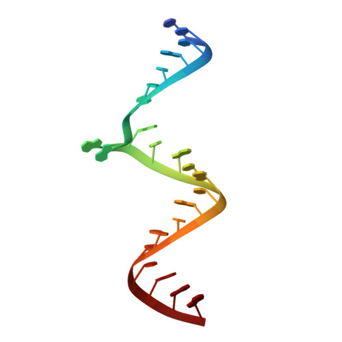
Cation-dependent cleavage of the duplex form of the subtype-B HIV-1 RNA dimerization initiation site.

Ennifar, E.Walter, P.Dumas, P.

(2010) Nucleic Acids Res 38: 5807-5816

  • DOI: https://doi.org/10.1093/nar/gkq344
  • Primary Citation of Related Structures:  
    2OIY, 2OJ0

  • PubMed Abstract: 

    The crystal structure of subtype-B HIV-1 genomic RNA Dimerization Initiation Site duplex revealed chain cleavage at a specific position resulting in 3'-phosphate and 5'-hydroxyl termini. A crystallographic analysis showed that Ba(2+), Mn(2+), Co(2+) and Zn(2+) bind specifically on a guanine base close to the cleaved position. The crystal structures also point to a necessary conformational change to induce an 'in-line' geometry at the cleavage site. In solution, divalent cations increased the rate of cleavage with pH/pKa compensation, indicating that a cation-bound hydroxide anion is responsible for the cleavage. We propose a 'Trojan horse' mechanism, possibly of general interest, wherein a doubly charged cation hosted near the cleavage site as a 'harmless' species is further transformed in situ into an 'aggressive' species carrying a hydroxide anion.


  • Organizational Affiliation

    Architecture et Réactivité de l'ARN, Université de Strasbourg, Institut de Biologie Moléculaire et Cellulaire du CNRS, 15 rue René Descartes F-67084 Strasbourg, France.


Macromolecules

Find similar nucleic acids by:  Sequence   |   3D Structure  

Entity ID: 1
MoleculeChains LengthOrganismImage
5'-R(*CP*UP*UP*GP*CP*UP*GP*AP*AP*GP*CP*GP*CP*GP*CP*AP*CP*GP*GP*CP*AP*AP*G)-3'
A, B
23N/A
Sequence Annotations
Expand
  • Reference Sequence
Experimental Data & Validation

Experimental Data

  • Method: X-RAY DIFFRACTION
  • Resolution: 1.60 Å
  • R-Value Free: 0.250 
  • R-Value Work: 0.242 
  • R-Value Observed: 0.242 
  • Space Group: P 21 21 2
Unit Cell:
Length ( Å )Angle ( ˚ )
a = 43.963α = 90
b = 47.55β = 90
c = 57.633γ = 90
Software Package:
Software NamePurpose
SHARPphasing
CNSrefinement
DENZOdata reduction
SCALEPACKdata scaling

Structure Validation

View Full Validation Report


Entry History 

Deposition Data

Revision History  (Full details and data files)

  • Version 1.0: 2007-12-25
    Type: Initial release
  • Version 1.1: 2011-07-13
    Changes: Version format compliance
  • Version 1.2: 2012-04-18
    Changes: Database references
  • Version 1.3: 2023-12-27
    Changes: Data collection, Database references, Derived calculations