2NTE

Crystal Structure of the BARD1 BRCT Domains


Experimental Data Snapshot

  • Method: X-RAY DIFFRACTION
  • Resolution: 1.90 Å
  • R-Value Free: 0.247 
  • R-Value Work: 0.196 
  • R-Value Observed: 0.199 

wwPDB Validation   3D Report Full Report


This is version 1.3 of the entry. See complete history


Literature

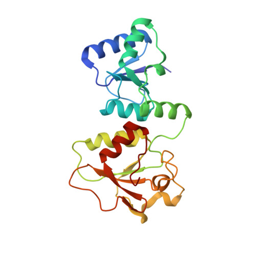
Crystal structure of the BARD1 BRCT domains.

Birrane, G.Varma, A.K.Soni, A.Ladias, J.A.

(2007) Biochemistry 46: 7706-7712

  • DOI: https://doi.org/10.1021/bi700323t
  • Primary Citation of Related Structures:  
    2NTE

  • PubMed Abstract: 

    The interaction of the breast tumor suppressor BRCA1 with the protein BARD1 results in the formation of a heterodimeric complex that has ubiquitin ligase activity and plays central roles in cell cycle checkpoint control and DNA repair. Both BRCA1 and BARD1 possess a pair of tandem BRCT domains that interact in a phosphorylation-dependent manner with target proteins. We determined the crystal structure of the human BARD1 BRCT repeats (residues 568-777) at 1.9 A resolution. The composition and structure of the BARD1 phosphoserine-binding pocket P1 are strikingly similar to those of the BRCA1 and MDC1 BRCT domains, suggesting a similar mode of interaction with the phosphate group of the ligand. By contrast, the BARD1 BRCT selectivity pocket P2 exhibits distinct structural features, including two prominent histidine residues, His685 and His686, which may be important for ligand binding. The protonation state of these histidines has a marked effect on the calculated electrostatic potential in the vicinity of P2, raising the possibility that ligand recognition may be regulated by changes in pH. Importantly, the BARD1 BRCT structure provides insights into the mechanisms by which the cancer-associated missense mutations C645R, V695L, and S761N may adversely affect the structure and function of BARD1.


  • Organizational Affiliation

    Molecular Medicine Laboratory and Macromolecular Crystallography Unit, Division of Experimental Medicine, Harvard Institutes of Medicine, Harvard Medical School, Boston, Massachusetts 02115, USA.


Macromolecules
Find similar proteins by:  (by identity cutoff)  |  3D Structure
Entity ID: 1
MoleculeChains Sequence LengthOrganismDetailsImage
BRCA1-associated RING domain protein 1
A, B
210Homo sapiensMutation(s): 0 
Gene Names: BARD1
UniProt & NIH Common Fund Data Resources
Find proteins for Q99728 (Homo sapiens)
Explore Q99728 
Go to UniProtKB:  Q99728
PHAROS:  Q99728
GTEx:  ENSG00000138376 
Entity Groups  
Sequence Clusters30% Identity50% Identity70% Identity90% Identity95% Identity100% Identity
UniProt GroupQ99728
Sequence Annotations
Expand
  • Reference Sequence
Small Molecules
Ligands 3 Unique
IDChains Name / Formula / InChI Key2D Diagram3D Interactions
SO4
Query on SO4

Download Ideal Coordinates CCD File 
C [auth A],
H [auth B]
SULFATE ION
O4 S
QAOWNCQODCNURD-UHFFFAOYSA-L
EDO
Query on EDO

Download Ideal Coordinates CCD File 
E [auth A],
F [auth A],
G [auth A],
K [auth B],
L [auth B]
1,2-ETHANEDIOL
C2 H6 O2
LYCAIKOWRPUZTN-UHFFFAOYSA-N
CL
Query on CL

Download Ideal Coordinates CCD File 
D [auth A],
I [auth B],
J [auth B]
CHLORIDE ION
Cl
VEXZGXHMUGYJMC-UHFFFAOYSA-M
Experimental Data & Validation

Experimental Data

  • Method: X-RAY DIFFRACTION
  • Resolution: 1.90 Å
  • R-Value Free: 0.247 
  • R-Value Work: 0.196 
  • R-Value Observed: 0.199 
  • Space Group: P 21 21 21
Unit Cell:
Length ( Å )Angle ( ˚ )
a = 57.441α = 90
b = 75.797β = 90
c = 116.57γ = 90
Software Package:
Software NamePurpose
REFMACrefinement
ADSCdata collection
HKL-2000data reduction
HKL-2000data scaling
SHELXSphasing

Structure Validation

View Full Validation Report


Entry History 

Deposition Data

Revision History  (Full details and data files)

  • Version 1.0: 2007-06-12
    Type: Initial release
  • Version 1.1: 2008-05-01
    Changes: Version format compliance
  • Version 1.2: 2011-07-13
    Changes: Advisory, Version format compliance
  • Version 1.3: 2023-12-27
    Changes: Data collection, Database references, Derived calculations