2NPZ

Crystal structure of junctioned hairpin ribozyme incorporating synthetic propyl linker


Experimental Data Snapshot

  • Method: X-RAY DIFFRACTION
  • Resolution: 3.35 Å
  • R-Value Free: 0.277 
  • R-Value Work: 0.261 
  • R-Value Observed: 0.268 

wwPDB Validation   3D Report Full Report


Ligand Structure Quality Assessment 


This is version 1.3 of the entry. See complete history


Literature

A posteriori design of crystal contacts to improve the X-ray diffraction properties of a small RNA enzyme.

MacElrevey, C.Spitale, R.C.Krucinska, J.Wedekind, J.E.

(2007) Acta Crystallogr D Biol Crystallogr 63: 812-825

  • DOI: https://doi.org/10.1107/S090744490702464X
  • Primary Citation of Related Structures:  
    2NPY, 2NPZ

  • PubMed Abstract: 

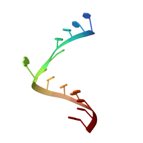
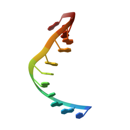
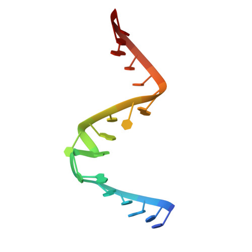
    The hairpin ribozyme is a small catalytic RNA comprising two helix-loop-helix domains linked by a four-way helical junction (4WJ). In its most basic form, each domain can be formed independently and reconstituted without a 4WJ to yield an active enzyme. The production of such minimal junctionless hairpin ribozymes is achievable by chemical synthesis, which has allowed structures to be determined for numerous nucleotide variants. However, abasic and other destabilizing core modifications hinder crystallization. This investigation describes the use of a dangling 5'-U to form an intermolecular U.U mismatch, as well as the use of synthetic linkers to tether the loop A and B domains, including (i) a three-carbon propyl linker (C3L) and (ii) a nine-atom triethylene glycol linker (S9L). Both linker constructs demonstrated similar enzymatic activity, but S9L constructs yielded crystals that diffracted to 2.65 A resolution or better. In contrast, C3L variants diffracted to 3.35 A and exhibited a 15 A expansion of the c axis. Crystal packing of the C3L construct showed a paucity of 6(1) contacts, which comprise numerous backbone to 2'-OH hydrogen bonds in junctionless and S9L complexes. Significantly, the crystal packing in minimal structures mimics stabilizing features observed in the 4WJ hairpin ribozyme structure. The results demonstrate how knowledge-based design can be used to improve diffraction and overcome otherwise destabilizing defects.


  • Organizational Affiliation

    Department of Biochemistry and Biophysics, University of Rochester School of Medicine and Dentistry, Rochester, New York 14642, USA.


Macromolecules

Find similar nucleic acids by:  Sequence   |   3D Structure  

Entity ID: 1
MoleculeChains LengthOrganismImage
5'-R(*UP*CP*CP*CP*AP*GP*UP*CP*CP*AP*CP*CP*G)-3'13N/A
Sequence Annotations
Expand
  • Reference Sequence

Find similar nucleic acids by:  Sequence   |   3D Structure  

Entity ID: 2
MoleculeChains LengthOrganismImage
5'-R(*CP*GP*GP*UP*GP*AP*GP*AP*AP*GP*GP*G)-3'12N/A
Sequence Annotations
Expand
  • Reference Sequence

Find similar nucleic acids by:  Sequence   |   3D Structure  

Entity ID: 3
MoleculeChains LengthOrganismImage
5'-R(*GP*GP*CP*AP*GP*AP*GP*AP*AP*AP*CP*AP*CP*AP*CP*GP*A)-3'17N/A
Sequence Annotations
Expand
  • Reference Sequence

Find similar nucleic acids by:  Sequence   |   3D Structure  

Entity ID: 4
MoleculeChains LengthOrganismImage
5'-R(*UP*CP*GP*UP*GP*GP*UP*AP*CP*AP*UP*UP*AP*CP*CP*UP*GP*CP*C)-3'19N/A
Sequence Annotations
Expand
  • Reference Sequence
Small Molecules
Ligands 2 Unique
IDChains Name / Formula / InChI Key2D Diagram3D Interactions
NCO
Query on NCO

Download Ideal Coordinates CCD File 
F [auth C]COBALT HEXAMMINE(III)
Co H18 N6
DYLMFCCYOUSRTK-UHFFFAOYSA-N
PDI
Query on PDI

Download Ideal Coordinates CCD File 
E [auth B]PHOSPHORIC ACID MONO-(3-HYDROXY-PROPYL) ESTER
C3 H9 O5 P
HYCSHFLKPSMPGO-UHFFFAOYSA-N
Experimental Data & Validation

Experimental Data

  • Method: X-RAY DIFFRACTION
  • Resolution: 3.35 Å
  • R-Value Free: 0.277 
  • R-Value Work: 0.261 
  • R-Value Observed: 0.268 
  • Space Group: P 61 2 2
Unit Cell:
Length ( Å )Angle ( ˚ )
a = 91.13α = 90
b = 91.13β = 90
c = 144.96γ = 120
Software Package:
Software NamePurpose
CNSrefinement
CrystalCleardata collection
CrystalCleardata reduction
CrystalCleardata scaling
CNSphasing

Structure Validation

View Full Validation Report


Ligand Structure Quality Assessment 


Entry History 

Deposition Data

Revision History  (Full details and data files)

  • Version 1.0: 2007-08-14
    Type: Initial release
  • Version 1.1: 2011-07-13
    Changes: Version format compliance
  • Version 1.2: 2023-12-27
    Changes: Data collection, Database references, Derived calculations
  • Version 1.3: 2024-04-03
    Changes: Refinement description