Structure of micelle-bound adrenomedullin: a first step toward the analysis of its interactions with receptors and small molecules.
Perez-Castells, J., Martin-Santamaria, S., Nieto, L., Ramos, A., Martinez, A., Pascual-Teresa, B., Jimenez-Barbero, J.(2012) Biopolymers 97: 45-53
- PubMed: 21830197
- DOI: https://doi.org/10.1002/bip.21700
- Primary Citation of Related Structures:
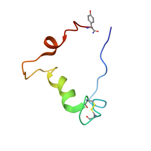
2L7S - PubMed Abstract:
Adrenomedullin (AM) is a regulatory peptide which plays many physiological roles including vasodilatation, bronchodilatation, hormone secretion regulation, growth, apoptosis, angiogenesis, and antimicrobial activities, among others. These regulatory activities make AM a relevant player in the pathophysiology of important diseases such as cardiovascular and renal conditions, cancer, and diabetes. Therefore, molecules that target the AM system have been proposed as having therapeutic potential. To guide the design and characterization of such molecules, we elucidated the three-dimensional structure of AM in a membrane mimicking medium using NMR spectroscopy methods. Under the employed experimental conditions, the structure can be described as composed by a central α-helical region, spanning about one third of its total length, flanked by two disordered segments at both N- and C-termini. The structure of AM in water is completely disordered. The 22-34 region of AM has a general tendency to adopt a helical structure under the employed experimental conditions. Furthermore, the study of the interaction of AM with two of its modulators has also been performed by using chemical shift perturbation analysis NMR methods with two-dimensional (2D)-TOCSY experiments, assisted with molecular modeling protocols. We expect these results will help in better understanding the interactions of AM with its receptor and binding proteins/molecules and in the development of novel modulators of AM activities.
Organizational Affiliation:
Departamento de Química, Facultad de Farmacia, Universidad San Pablo, Madrid, Spain.