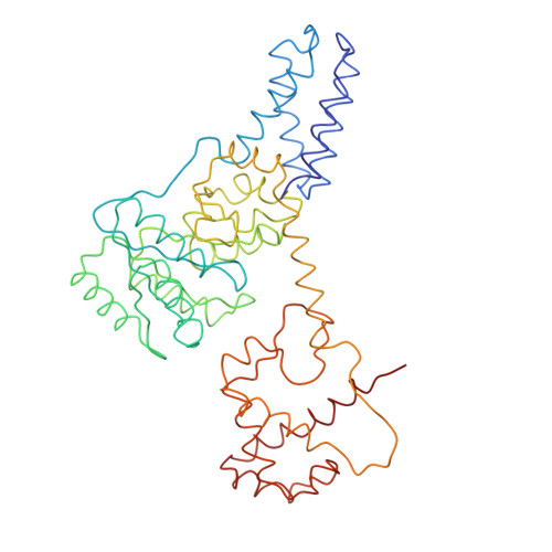
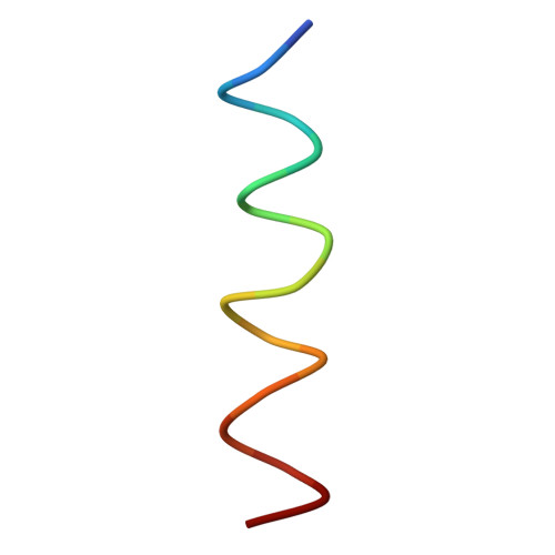
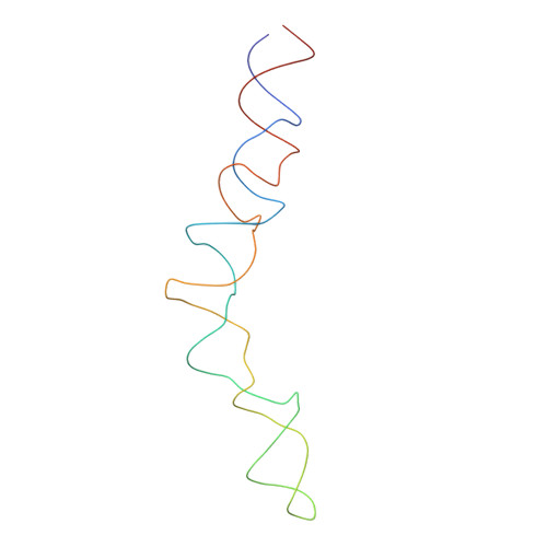
2IY3

Structure of the E. Coli Signal Regognition Particle


Experimental Data Snapshot

  • Method: ELECTRON MICROSCOPY
  • Resolution: 16.0 Å
  • Aggregation State: PARTICLE 
  • Reconstruction Method: SINGLE PARTICLE 

wwPDB Validation   3D Report Full Report


This is version 1.3 of the entry. See complete history


Literature

Structure of the E. Coli Signal Recognition Particle Bound to a Translating Ribosome

Schaffitzel, C.Oswald, M.Berger, I.Ishikawa, T.Abrahams, J.P.Koerten, H.K.Koning, R.I.Ban, N.

(2006) Nature 444: 503

  • DOI: https://doi.org/10.1038/nature05182
  • Primary Citation of Related Structures:  
    2IY3

  • PubMed Abstract: 

    The prokaryotic signal recognition particle (SRP) targets membrane proteins into the inner membrane. It binds translating ribosomes and screens the emerging nascent chain for a hydrophobic signal sequence, such as the transmembrane helix of inner membrane proteins. If such a sequence emerges, the SRP binds tightly, allowing the SRP receptor to lock on. This assembly delivers the ribosome-nascent chain complex to the protein translocation machinery in the membrane. Using cryo-electron microscopy and single-particle reconstruction, we obtained a 16 A structure of the Escherichia coli SRP in complex with a translating E. coli ribosome containing a nascent chain with a transmembrane helix anchor. We also obtained structural information on the SRP bound to an empty E. coli ribosome. The latter might share characteristics with a scanning SRP complex, whereas the former represents the next step: the targeting complex ready for receptor binding. High-resolution structures of the bacterial ribosome and of the bacterial SRP components are available, and their fitting explains our electron microscopic density. The structures reveal the regions that are involved in complex formation, provide insight into the conformation of the SRP on the ribosome and indicate the conformational changes that accompany high-affinity SRP binding to ribosome nascent chain complexes upon recognition of the signal sequence.


  • Organizational Affiliation

    ETH Zurich, Institute for Molecular Biology and Biophysics, HPK Building, Schafmattstrasse 20, 8093 Zurich, Switzerland.


Macromolecules

Find similar proteins by:  (by identity cutoff)  |  3D Structure
Entity ID: 1
MoleculeChains Sequence LengthOrganismDetailsImage
Signal recognition particle protein,Signal recognition particle 54 kDa protein432Thermus aquaticusSaccharolobus solfataricus
This entity is chimeric
Mutation(s): 0 
Gene Names: ffhsrp54SULA_1982SULB_1983SULC_1981
UniProt
Find proteins for Q97ZE7 (Saccharolobus solfataricus (strain ATCC 35092 / DSM 1617 / JCM 11322 / P2))
Explore Q97ZE7 
Go to UniProtKB:  Q97ZE7
Find proteins for O07347 (Thermus aquaticus)
Explore O07347 
Go to UniProtKB:  O07347
Entity Groups  
Sequence Clusters30% Identity50% Identity70% Identity90% Identity95% Identity100% Identity
UniProt GroupsQ97ZE7O07347
Sequence Annotations
Expand
  • Reference Sequence

Find similar proteins by:  Sequence   |   3D Structure  

Entity ID: 3
MoleculeChains Sequence LengthOrganismDetailsImage
SIGNAL SEQUENCE17synthetic constructMutation(s): 0 
Entity Groups  
Sequence Clusters30% Identity50% Identity70% Identity90% Identity95% Identity100% Identity
Sequence Annotations
Expand
  • Reference Sequence
Find similar nucleic acids by:  (by identity cutoff)  |  3D Structure
Entity ID: 2
MoleculeChains LengthOrganismImage
4.5S RNA110Escherichia coli
Sequence Annotations
Expand
  • Reference Sequence
Experimental Data & Validation

Experimental Data

  • Method: ELECTRON MICROSCOPY
  • Resolution: 16.0 Å
  • Aggregation State: PARTICLE 
  • Reconstruction Method: SINGLE PARTICLE 
EM Software:
TaskSoftware PackageVersion
RECONSTRUCTIONSPIDER

Structure Validation

View Full Validation Report


Entry History 

Deposition Data

Revision History  (Full details and data files)

  • Version 1.0: 2006-11-02
    Type: Initial release
  • Version 1.1: 2013-08-07
    Changes: Derived calculations, Other, Refinement description, Version format compliance
  • Version 1.2: 2017-03-15
    Changes: Source and taxonomy
  • Version 1.3: 2017-08-23
    Changes: Data collection, Refinement description