Pathogenicity of the BRCA1 missense variant M1775K is determined by the disruption of the BRCT phosphopeptide-binding pocket: a multi-modal approach.
Tischkowitz, M., Hamel, N., Carvalho, M.A., Birrane, G., Soni, A., van Beers, E.H., Joosse, S.A., Wong, N., Novak, D., Quenneville, L.A., Grist, S.A., Nederlof, P.M., Goldgar, D.E., Tavtigian, S.V., Monteiro, A.N., Ladias, J.A., Foulkes, W.D.(2008) Eur J Hum Genet 16: 820-832
- PubMed: 18285836
- DOI: https://doi.org/10.1038/ejhg.2008.13
- Primary Citation of Related Structures:
2ING - PubMed Abstract:
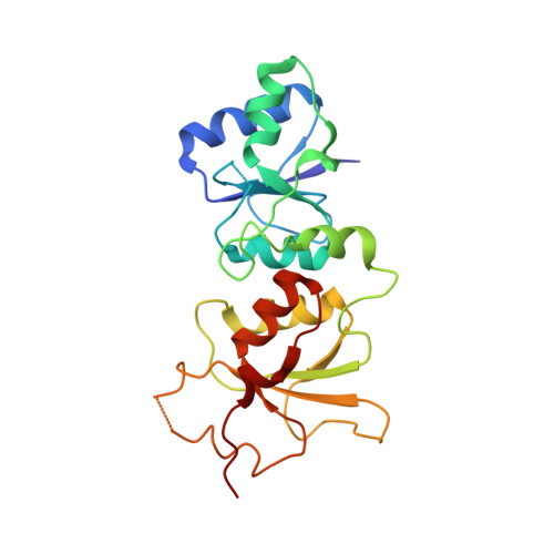
A number of germ-line mutations in the BRCA1 gene confer susceptibility to breast and ovarian cancer. However, it remains difficult to determine whether many single amino-acid (missense) changes in the BRCA1 protein that are frequently detected in the clinical setting are pathologic or not. Here, we used a combination of functional, crystallographic, biophysical, molecular and evolutionary techniques, and classical genetic segregation analysis to demonstrate that the BRCA1 missense variant M1775K is pathogenic. Functional assays in yeast and mammalian cells showed that the BRCA1 BRCT domains carrying the amino-acid change M1775K displayed markedly reduced transcriptional activity, indicating that this variant represents a deleterious mutation. Importantly, the M1775K mutation disrupted the phosphopeptide-binding pocket of the BRCA1 BRCT domains, thereby inhibiting the BRCA1 interaction with the proteins BRIP1 and CtIP, which are involved in DNA damage-induced checkpoint control. These results indicate that the integrity of the BRCT phosphopeptide-binding pocket is critical for the tumor suppression function of BRCA1. Moreover, this study demonstrates that multiple lines of evidence obtained from a combination of functional, structural, molecular and evolutionary techniques, and classical genetic segregation analysis are required to confirm the pathogenicity of rare variants of disease-susceptibility genes and obtain important insights into the underlying pathogenetic mechanisms.
Organizational Affiliation:
Department of Oncology, McGill University, Montréal, Quebec, Canada.