2HVY

Crystal structure of an H/ACA box RNP from Pyrococcus furiosus


Experimental Data Snapshot

  • Method: X-RAY DIFFRACTION
  • Resolution: 2.30 Å
  • R-Value Free: 0.278 
  • R-Value Work: 0.238 
  • R-Value Observed: 0.240 

wwPDB Validation   3D Report Full Report


Ligand Structure Quality Assessment 


This is version 1.4 of the entry. See complete history


Literature

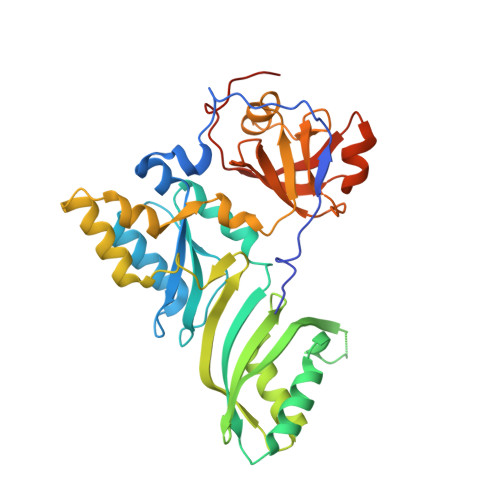
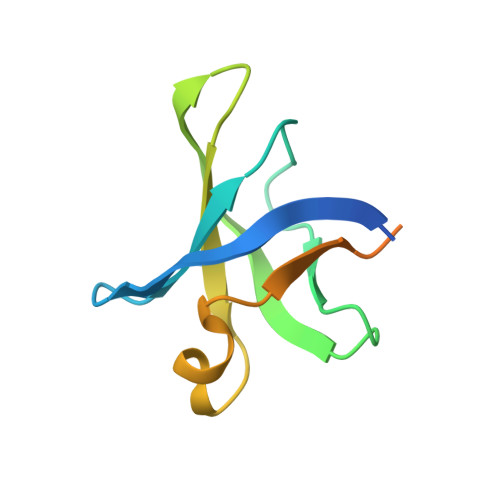
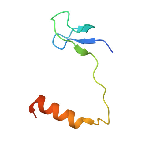
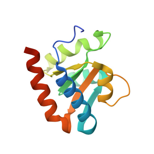
Crystal structure of an H/ACA box ribonucleoprotein particle

Li, L.Ye, K.

(2006) Nature 443: 302-307

  • DOI: https://doi.org/10.1038/nature05151
  • Primary Citation of Related Structures:  
    2HVY

  • PubMed Abstract: 

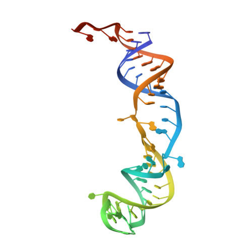
    H/ACA ribonucleoprotein particles (RNPs) are a family of RNA pseudouridine synthases that specify modification sites through guide RNAs. They also participate in eukaryotic ribosomal RNA processing and are a component of vertebrate telomerases. Here we report the crystal structure, at 2.3 A resolution, of an entire archaeal H/ACA RNP consisting of proteins Cbf5, Nop10, Gar1 and L7ae, and a single-hairpin H/ACA RNA, revealing a modular organization of the complex. The RNA upper stem is bound to a composite surface formed by L7ae, Nop10 and Cbf5, and the RNA lower stem and ACA signature motif are bound to the PUA domain of Cbf5, thereby positioning middle guide sequences so that they are primed to pair with substrate RNA. Furthermore, Gar1 may regulate substrate loading and release. The structure rationalizes the consensus structure of H/ACA RNAs, suggests a functional role of each protein, and provides a framework for understanding the mechanism of RNA-guided pseudouridylation, as well as various cellular functions of H/ACA RNP.


  • Organizational Affiliation

    National Institute of Biological Sciences, 7 Science Park Road, Zhongguancun Life Science Park, Beijing 102206, China.


Macromolecules

Find similar proteins by:  (by identity cutoff)  |  3D Structure
Entity ID: 2
MoleculeChains Sequence LengthOrganismDetailsImage
Probable tRNA pseudouridine synthase BB [auth A]346Pyrococcus furiosusMutation(s): 0 
Gene Names: truB
EC: 5.4.99
UniProt
Find proteins for Q7LWY0 (Pyrococcus furiosus (strain ATCC 43587 / DSM 3638 / JCM 8422 / Vc1))
Explore Q7LWY0 
Go to UniProtKB:  Q7LWY0
Entity Groups  
Sequence Clusters30% Identity50% Identity70% Identity90% Identity95% Identity100% Identity
UniProt GroupQ7LWY0
Sequence Annotations
Expand
  • Reference Sequence
Find similar proteins by:  (by identity cutoff)  |  3D Structure
Entity ID: 3
MoleculeChains Sequence LengthOrganismDetailsImage
Small nucleolar rnp similar to gar1C [auth B]104Pyrococcus furiosusMutation(s): 0 
Gene Names: GAR1
UniProt
Find proteins for Q8U029 (Pyrococcus furiosus (strain ATCC 43587 / DSM 3638 / JCM 8422 / Vc1))
Explore Q8U029 
Go to UniProtKB:  Q8U029
Entity Groups  
Sequence Clusters30% Identity50% Identity70% Identity90% Identity95% Identity100% Identity
UniProt GroupQ8U029
Sequence Annotations
Expand
  • Reference Sequence
Find similar proteins by:  (by identity cutoff)  |  3D Structure
Entity ID: 4
MoleculeChains Sequence LengthOrganismDetailsImage
Ribosome biogenesis protein Nop10D [auth C]60Pyrococcus furiosusMutation(s): 1 
Gene Names: NOP10
UniProt
Find proteins for Q8U1R4 (Pyrococcus furiosus (strain ATCC 43587 / DSM 3638 / JCM 8422 / Vc1))
Explore Q8U1R4 
Go to UniProtKB:  Q8U1R4
Entity Groups  
Sequence Clusters30% Identity50% Identity70% Identity90% Identity95% Identity100% Identity
UniProt GroupQ8U1R4
Sequence Annotations
Expand
  • Reference Sequence
Find similar proteins by:  (by identity cutoff)  |  3D Structure
Entity ID: 5
MoleculeChains Sequence LengthOrganismDetailsImage
50S ribosomal protein L7AeE [auth D]130Pyrococcus furiosusMutation(s): 1 
Gene Names: rpl7ae
UniProt
Find proteins for Q8U160 (Pyrococcus furiosus (strain ATCC 43587 / DSM 3638 / JCM 8422 / Vc1))
Explore Q8U160 
Go to UniProtKB:  Q8U160
Entity Groups  
Sequence Clusters30% Identity50% Identity70% Identity90% Identity95% Identity100% Identity
UniProt GroupQ8U160
Sequence Annotations
Expand
  • Reference Sequence
Find similar nucleic acids by:  (by identity cutoff)  |  3D Structure
Entity ID: 1
MoleculeChains LengthOrganismImage
H/ACA RNAA [auth E]65N/A
Sequence Annotations
Expand
  • Reference Sequence
Small Molecules
Ligands 2 Unique
IDChains Name / Formula / InChI Key2D Diagram3D Interactions
ATP
Query on ATP

Download Ideal Coordinates CCD File 
F [auth A]ADENOSINE-5'-TRIPHOSPHATE
C10 H16 N5 O13 P3
ZKHQWZAMYRWXGA-KQYNXXCUSA-N
ZN
Query on ZN

Download Ideal Coordinates CCD File 
G [auth C]ZINC ION
Zn
PTFCDOFLOPIGGS-UHFFFAOYSA-N
Experimental Data & Validation

Experimental Data

  • Method: X-RAY DIFFRACTION
  • Resolution: 2.30 Å
  • R-Value Free: 0.278 
  • R-Value Work: 0.238 
  • R-Value Observed: 0.240 
  • Space Group: P 21 21 21
Unit Cell:
Length ( Å )Angle ( ˚ )
a = 83.031α = 90
b = 90.95β = 90
c = 114.064γ = 90
Software Package:
Software NamePurpose
REFMACrefinement
HKL-2000data reduction
HKL-2000data scaling
MOLREPphasing

Structure Validation

View Full Validation Report


Ligand Structure Quality Assessment 


Entry History 

Deposition Data

  • Released Date: 2006-09-12 
  • Deposition Author(s): Ye, K.

Revision History  (Full details and data files)

  • Version 1.0: 2006-09-12
    Type: Initial release
  • Version 1.1: 2008-05-01
    Changes: Version format compliance
  • Version 1.2: 2011-07-13
    Changes: Advisory, Version format compliance
  • Version 1.3: 2021-11-10
    Changes: Database references, Derived calculations
  • Version 1.4: 2023-10-25
    Changes: Data collection, Refinement description