2GTT

Crystal structure of the rabies virus nucleoprotein-RNA complex


Experimental Data Snapshot

  • Method: X-RAY DIFFRACTION
  • Resolution: 3.49 Å
  • R-Value Free: 0.255 
  • R-Value Work: 0.224 
  • R-Value Observed: 0.225 

wwPDB Validation   3D Report Full Report


This is version 2.0 of the entry. See complete history


Literature

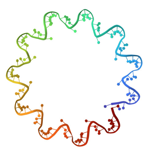
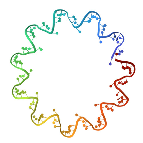
Crystal Structure of the Rabies Virus Nucleoprotein-RNA Complex

Albertini, A.A.V.Wernimont, A.K.Muziol, T.Ravelli, R.B.G.Clapier, C.R.Schoehn, G.Weissenhorn, W.Ruigrok, R.W.H.

(2006) Science 313: 360-363

  • DOI: https://doi.org/10.1126/science.1125280
  • Primary Citation of Related Structures:  
    2GTT

  • PubMed Abstract: 

    Negative-strand RNA viruses condense their genome into a helical nucleoprotein-RNA complex, the nucleocapsid, which is packed into virions and serves as a template for the RNA-dependent RNA polymerase complex. The crystal structure of a recombinant rabies virus nucleoprotein-RNA complex, organized in an undecameric ring, has been determined at 3.5 angstrom resolution. Polymerization of the nucleoprotein is achieved by domain exchange between protomers, with flexible hinges allowing nucleocapsid formation. The two core domains of the nucleoprotein clamp around the RNA at their interface and shield it from the environment. RNA sequestering by nucleoproteins is likely a common mechanism used by negative-strand RNA viruses to protect their genomes from the innate immune response directed against viral RNA in human host cells at certain stages of an infectious cycle.


  • Organizational Affiliation

    Institut de Virologie Moléculaire et Structurale, FRE 2854 Université Joseph Fourier-CNRS, Boite Postale 181, 38042 Grenoble, France.


Macromolecules

Find similar proteins by:  (by identity cutoff)  |  3D Structure
Entity ID: 1
MoleculeChains Sequence LengthOrganismDetailsImage
Nucleoprotein
A, B, C, D, E
A, B, C, D, E, F, G, H, I, J, K, L, M, N, O, P, Q, R, S, T, U, V
450Lyssavirus rabiesMutation(s): 0 
UniProt
Find proteins for P0DOF3 (Rabies virus (strain ERA))
Explore P0DOF3 
Go to UniProtKB:  P0DOF3
Entity Groups  
Sequence Clusters30% Identity50% Identity70% Identity90% Identity95% Identity100% Identity
UniProt GroupP0DOF3
Sequence Annotations
Expand
  • Reference Sequence
Find similar nucleic acids by:  (by identity cutoff)  |  3D Structure
Entity ID: 2
MoleculeChains LengthOrganismImage
RNA (99-MER)99synthetic construct
Sequence Annotations
Expand
  • Reference Sequence
Find similar nucleic acids by:  (by identity cutoff)  |  3D Structure
Entity ID: 3
MoleculeChains LengthOrganismImage
RNA (99-MER)99synthetic construct
Sequence Annotations
Expand
  • Reference Sequence
Experimental Data & Validation

Experimental Data

  • Method: X-RAY DIFFRACTION
  • Resolution: 3.49 Å
  • R-Value Free: 0.255 
  • R-Value Work: 0.224 
  • R-Value Observed: 0.225 
  • Space Group: P 21 21 2
Unit Cell:
Length ( Å )Angle ( ˚ )
a = 270.43α = 90
b = 281β = 90
c = 236.9γ = 90
Software Package:
Software NamePurpose
PHENIXrefinement
ProDCdata collection
XDSdata scaling
SHELXDphasing

Structure Validation

View Full Validation Report


Entry History 

Deposition Data

Revision History  (Full details and data files)

  • Version 1.0: 2006-09-19
    Type: Initial release
  • Version 1.1: 2008-05-01
    Changes: Version format compliance
  • Version 1.2: 2011-07-13
    Changes: Advisory, Version format compliance
  • Version 1.3: 2017-10-18
    Changes: Refinement description
  • Version 2.0: 2023-10-18
    Type: Coordinate replacement
    Reason: Polymer backbone linkage
    Changes: Advisory, Atomic model, Data collection, Database references, Derived calculations, Non-polymer description, Other, Polymer sequence, Refinement description, Source and taxonomy, Structure summary