NMR Structure of the Full-length Linear Dimer of Stem-Loop-1 RNA in the HIV-1 Dimer Initiation Site.
Ulyanov, N.B., Mujeeb, A., Du, Z., Tonelli, M., Parslow, T.G., James, T.L.(2006) J Biol Chem 281: 16168-16177
- PubMed: 16603544
- DOI: https://doi.org/10.1074/jbc.M601711200
- Primary Citation of Related Structures:
2GM0 - PubMed Abstract:
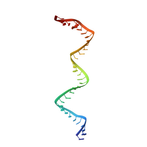
The packaging signal of HIV-1 RNA contains a stem-loop structure, SL1, which serves as the dimerization initiation site for two identical copies of the genome and is important for packaging of the RNA genome into the budding virion and for overall infectivity. SL1 spontaneously dimerizes via a palindromic hexanucleotide sequence in its apical loop, forming a metastable kissing dimer form. Incubation with nucleocapsid protein causes this form to refold to a thermodynamically stable mature linear dimer. Here, we present an NMR structure of the latter form of the full-length SL1 sequence of the Lai HIV-1 isolate. The structure was refined using nuclear Overhauser effect and residual dipolar coupling data. The structure presents a symmetric homodimer of two RNA strands of 35 nucleotides each; it includes five stems separated by four internal loops. The central palindromic stem is surrounded by two symmetric adenine-rich 1-2 internal loops, A-bulges. All three adenines in each A-bulge are stacked inside the helix, consistent with the solution structures of shorter SL1 constructs determined previously. The outer 4-base pair stems and, proximal to them, purine-rich 1-3 internal loops, or G-bulges, are the least stable parts of the molecule. The G-bulges display high conformational variability in the refined ensemble of structures, despite the availability of many structural restraints for this region. Nevertheless, most conformations share a similar structural motif: a guanine and an adenine from opposite strands form a GA mismatch stacked on the top of the neighboring stem. The two remaining guanines are exposed, one in the minor groove and another in the major groove side of the helix, consistent with secondary structure probing data for SL1. These guanines may be recognized by the nucleocapsid protein, which binds tightly to the G-bulge in vitro.
Organizational Affiliation:
Department of Pharmaceutical Chemistry, University of California, San Francisco, California 94143, USA.