2FQN

Crystal structure of the Homo sapiens cytoplasmic ribosomal decoding A site


Experimental Data Snapshot

  • Method: X-RAY DIFFRACTION
  • Resolution: 2.30 Å
  • R-Value Free: 0.282 
  • R-Value Work: 0.247 

wwPDB Validation   3D Report Full Report


This is version 1.3 of the entry. See complete history


Literature

Two conformational states in the crystal structure of the Homo sapiens cytoplasmic ribosomal decoding A site.

Kondo, J.Urzhumtsev, A.Westhof, E.

(2006) Nucleic Acids Res 34: 676-685

  • DOI: https://doi.org/10.1093/nar/gkj467
  • Primary Citation of Related Structures:  
    2FQN

  • PubMed Abstract: 

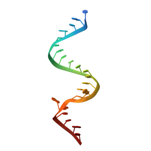
    The decoding A site of the small ribosomal subunit is an RNA molecular switch, which monitors codon-anticodon interactions to guarantee translation fidelity. We have solved the crystal structure of an RNA fragment containing two Homo sapiens cytoplasmic A sites. Each of the two A sites presents a different conformational state. In one state, adenines A1492 and A1493 are fully bulged-out with C1409 forming a wobble-like pair to A1491. In the second state, adenines A1492 and A1493 form non-Watson-Crick pairs with C1409 and G1408, respectively while A1491 bulges out. The first state of the eukaryotic A site is, thus, basically the same as in the bacterial A site with bulging A1492 and A1493. It is the state used for recognition of the codon/anticodon complex. On the contrary, the second state of the H.sapiens cytoplasmic A site is drastically different from any of those observed for the bacterial A site without bulging A1492 and A1493.


  • Organizational Affiliation

    Institut de Biologie Moléculaire et Cellulaire, UPR9002 CNRS, Université Louis Pasteur, 15 rue René Descartes, 67084 Strasbourg, France.


Macromolecules

Find similar nucleic acids by:  Sequence   |   3D Structure  

Entity ID: 1
MoleculeChains LengthOrganismImage
5'-R(*UP*UP*GP*CP*GP*UP*CP*GP*CP*UP*CP*CP*GP*GP*AP*AP*AP*AP*GP*UP*CP*GP*C)-3'
A, B
23N/A
Sequence Annotations
Expand
  • Reference Sequence
Experimental Data & Validation

Experimental Data

  • Method: X-RAY DIFFRACTION
  • Resolution: 2.30 Å
  • R-Value Free: 0.282 
  • R-Value Work: 0.247 
  • Space Group: P 21 21 2
Unit Cell:
Length ( Å )Angle ( ˚ )
a = 46.05α = 90
b = 47.39β = 90
c = 56.76γ = 90
Software Package:
Software NamePurpose
d*TREKdata processing
CNSrefinement
PDB_EXTRACTdata extraction
CrystalCleardata reduction
CrystalCleardata scaling
AMoREphasing

Structure Validation

View Full Validation Report


Entry History 

Deposition Data

Revision History  (Full details and data files)

  • Version 1.0: 2006-02-14
    Type: Initial release
  • Version 1.1: 2008-05-01
    Changes: Version format compliance
  • Version 1.2: 2011-07-13
    Changes: Version format compliance
  • Version 1.3: 2024-02-14
    Changes: Data collection, Database references, Derived calculations