2FE3

The crystal structure of bacillus subtilis PerR-Zn reveals a novel Zn(Cys)4 Structural redox switch


Experimental Data Snapshot

  • Method: X-RAY DIFFRACTION
  • Resolution: 1.75 Å
  • R-Value Free: 0.229 
  • R-Value Work: 0.172 
  • R-Value Observed: 0.172 

wwPDB Validation   3D Report Full Report


This is version 1.3 of the entry. See complete history


Literature

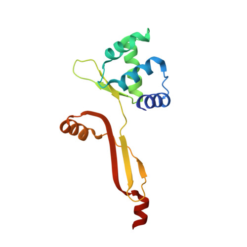
Crystal structure of the apo-PerR-Zn protein from Bacillus subtilis.

Traore, D.A.K.El Ghazouani, A.Ilango, S.Dupuy, J.Jacquamet, L.Ferrer, J.-L.Caux-Thang, C.Duarte, V.Latour, J.M.

(2006) Mol Microbiol 61: 1211-1219

  • DOI: https://doi.org/10.1111/j.1365-2958.2006.05313.x
  • Primary Citation of Related Structures:  
    2FE3

  • PubMed Abstract: 

    Bacteria adapt to elevated levels of Reactive Oxygen Species (ROS) by increasing the expression of defence and repair proteins, which is regulated by ROS responsive transcription factors. In Bacillus subtilis the zinc protein PerR, a peroxide sensor that binds DNA in the presence of a regulatory metal Mn2+ or Fe2+, mediates the adaptive response to H2O2. This study presents the first crystal structure of apo-PerR-Zn which shows that all four cysteine residues of the protein are involved in zinc co-ordination. The Zn(Cys)4 site locks the dimerization domain and stabilizes the dimer. Sequence alignment of PerR-like proteins supports that this structural site may constitute a distinctive feature of this class of peroxide stress regulators.


  • Organizational Affiliation

    DRDC/Laboratoire de Physicochimie des Métaux en Biologie, UMR 5155 CEA-CNRS-UJF, CEA-Grenoble, 38054 Grenoble Cedex 9, France.


Macromolecules
Find similar proteins by:  (by identity cutoff)  |  3D Structure
Entity ID: 1
MoleculeChains Sequence LengthOrganismDetailsImage
Peroxide operon regulator
A, B
145Bacillus subtilisMutation(s): 0 
UniProt
Find proteins for P71086 (Bacillus subtilis (strain 168))
Explore P71086 
Go to UniProtKB:  P71086
Entity Groups  
Sequence Clusters30% Identity50% Identity70% Identity90% Identity95% Identity100% Identity
UniProt GroupP71086
Sequence Annotations
Expand
  • Reference Sequence
Experimental Data & Validation

Experimental Data

  • Method: X-RAY DIFFRACTION
  • Resolution: 1.75 Å
  • R-Value Free: 0.229 
  • R-Value Work: 0.172 
  • R-Value Observed: 0.172 
  • Space Group: P 1
Unit Cell:
Length ( Å )Angle ( ˚ )
a = 41.29α = 82.18
b = 42.16β = 80.11
c = 53.49γ = 60.96
Software Package:
Software NamePurpose
REFMACrefinement
PDB_EXTRACTdata extraction
XDSdata scaling
SOLVEphasing

Structure Validation

View Full Validation Report


Entry History 

Deposition Data

Revision History  (Full details and data files)

  • Version 1.0: 2006-12-19
    Type: Initial release
  • Version 1.1: 2008-04-01
    Changes: Version format compliance
  • Version 1.2: 2011-07-13
    Changes: Derived calculations, Version format compliance
  • Version 1.3: 2024-02-14
    Changes: Data collection, Database references, Derived calculations