Analyses of Variant Acid beta-Glucosidases: EFFECTS OF GAUCHER DISEASE MUTATIONS.
Liou, B., Kazimierczuk, A., Zhang, M., Scott, C.R., Hegde, R.S., Grabowski, G.A.(2006) J Biol Chem 281: 4242-4253
- PubMed: 16293621
- DOI: https://doi.org/10.1074/jbc.M511110200
- Primary Citation of Related Structures:
2F61 - PubMed Abstract:
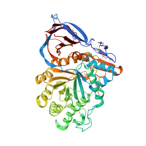
Acid beta-glucosidase (GCase) is a 497-amino acid, membrane-associated lysosomal exo-beta-glucosidase whose defective activity leads to the Gaucher disease phenotypes. To move toward a structure/function map for disease mutations, 52 selected single amino acid substitutions were introduced into GCase, expressed in an insect cell system, purified, and characterized for basic kinetic, stability, and activator response properties. The variant GCases from Gaucher disease patients and selected variant GCases from the mouse had decreased relative k(cat) and differential effects on active site binding and/or attachment of mechanism-based covalent (conduritol B epoxide) or reversible (deoxynojirimycin derivatives) inhibitors. A defect in negatively charged phospholipid activation was present in the majority of variant GCases but was increased in two, N370S and V394L. Deficits in saposin C enhancement of k(cat) were present in variant GCases involving residues 48-122, whereas approximately 2-fold increases were obtained with the L264I GCase. About 50% of variant GCases each had wild-type or increased sensitivity to in vitro cathepsin D digestion. Mapping of these properties onto the crystal structures of GCase indicated wide dispersion of functional properties that can affect catalytic function and stability. Site-directed mutagenesis of cysteine residues showed that the disulfide bonds, Cys(4)-Cys(16) and Cys(18)-Cys(23), and a free Cys(342) were essential for activity; the free Cys(126) and Cys(248) were not. Relative k(cat) was highly sensitive to a His substitution at Arg(496) but not at Arg(495). These studies and high phylogenetic conservation indicate localized and general structural effects of Gaucher disease mutations that were not obvious from the nature of the amino acid substitution, including those predicted to be nondisruptive (e.g. Val --> Leu). These results provide initial studies for the engineering of variant GCases and, potentially, molecular chaperones for therapeutic use.
Organizational Affiliation:
Division and Program in Human Genetics, Children's Hospital Research Foundation, Cincinnati, OH 45229, USA.