2ENR

CO-CRYSTALS OF DEMETALLIZED CONCANAVALIN A WITH CADMIUM HAVING A CADMIUM ION BOUND IN BOTH THE S1 SITE AND THE S2 SITE


Experimental Data Snapshot

  • Method: X-RAY DIFFRACTION
  • Resolution: 2.35 Å
  • R-Value Work: 0.173 
  • R-Value Observed: 0.173 

wwPDB Validation   3D Report Full Report


This is version 1.4 of the entry. See complete history


Literature

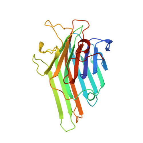
Zinc/calcium- and cadmium/cadmium-substituted concanavalin A: interplay of metal binding, pH and molecular packing.

Bouckaert, J.Loris, R.Wyns, L.

(2000) Acta Crystallogr D Biol Crystallogr 56: 1569-1576

  • DOI: https://doi.org/10.1107/s0907444900013342
  • Primary Citation of Related Structures:  
    2ENR, 3ENR

  • PubMed Abstract: 

    The crystal structures of cadmium/cadmium and zinc/calcium concanavalin A (con A) at pH 5.0 and pH 6.15, respectively, were determined. The structure of cadmium/cadmium con A confirms that the secondary Cd(2+)-binding site S3 is empty at pH 5. The metal-binding sites S1 and S2 are only very slightly affected by the substitution with cadmium. On the other hand, S1 and S2 and most of the protein surface of zinc/calcium con A at pH 6.15 differ from other fully metal-bound and carbohydrate-free structures. Most of these structural differences at the protein surface are a result of the interplay between metal binding, protonation and crystal packing. This interplay is expressed by relative rotations and translations of the con A units in alternative crystal packings and participation in space-group conversions inside crystals in situ. The particular crystal packing of zinc/calcium con A creates a novel zinc-binding site S4. The Zn(2+) ion in S4 ligates two aspartates from one tetramer and a histidine from a symmetry-related tetramer.


  • Organizational Affiliation

    Laboratorium voor Ultrastructuur, Vlaams Interuniversitair Instituut voor Biotechnologie, Vrije Universiteit Brussel, Paardenstraat 65, B-1640 Sint-Genesius-Rode, Belgium. bouckaej@vub.ac.be


Macromolecules
Find similar proteins by:  (by identity cutoff)  |  3D Structure
Entity ID: 1
MoleculeChains Sequence LengthOrganismDetailsImage
CONCANAVALIN A237Canavalia ensiformisMutation(s): 0 
UniProt
Find proteins for P02866 (Canavalia ensiformis)
Explore P02866 
Go to UniProtKB:  P02866
Entity Groups  
Sequence Clusters30% Identity50% Identity70% Identity90% Identity95% Identity100% Identity
UniProt GroupP02866
Sequence Annotations
Expand
  • Reference Sequence
Experimental Data & Validation

Experimental Data

  • Method: X-RAY DIFFRACTION
  • Resolution: 2.35 Å
  • R-Value Work: 0.173 
  • R-Value Observed: 0.173 
  • Space Group: I 2 2 2
Unit Cell:
Length ( Å )Angle ( ˚ )
a = 63.2α = 90
b = 87.69β = 90
c = 89.23γ = 90
Software Package:
Software NamePurpose
X-PLORmodel building
X-PLORrefinement
MADNESSdata reduction
CCP4data scaling
X-PLORphasing

Structure Validation

View Full Validation Report


Entry History 

Deposition Data

Revision History  (Full details and data files)

  • Version 1.0: 1999-02-16
    Type: Initial release
  • Version 1.1: 2008-03-24
    Changes: Version format compliance
  • Version 1.2: 2011-07-13
    Changes: Version format compliance
  • Version 1.3: 2018-04-18
    Changes: Data collection, Other
  • Version 1.4: 2023-08-09
    Changes: Database references, Derived calculations, Refinement description