2DQA

Crystal Structure of Tapes japonica Lysozyme


Experimental Data Snapshot

  • Method: X-RAY DIFFRACTION
  • Resolution: 1.60 Å
  • R-Value Free: 0.210 
  • R-Value Work: 0.175 
  • R-Value Observed: 0.176 

wwPDB Validation   3D Report Full Report


Ligand Structure Quality Assessment 


This is version 3.0 of the entry. See complete history


Literature

Crystal Structure of Tapes japonica Lysozyme with Substrate Analogue: STRUCTURAL BASIS OF THE CATALYTIC MECHANISM AND MANIFESTATION OF ITS CHITINASE ACTIVITY ACCOMPANIED BY QUATERNARY STRUCTURAL CHANGE

Goto, T.Abe, Y.Kakuta, Y.Takeshita, K.Imoto, T.Ueda, T.

(2007) J Biol Chem 282: 27459-27467

  • DOI: https://doi.org/10.1074/jbc.M704555200
  • Primary Citation of Related Structures:  
    2DQA

  • PubMed Abstract: 

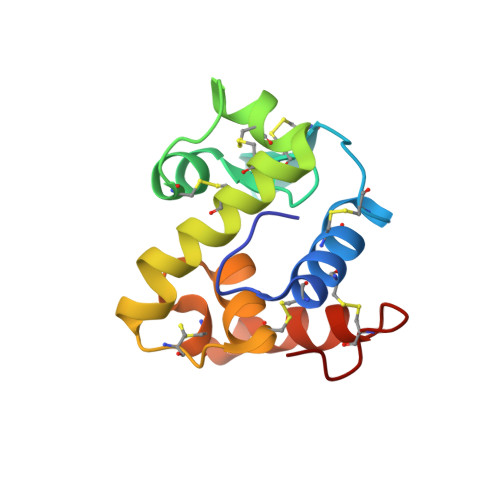
    Tapes japonica lysozyme (TJL) is classified as a member of the recently established i-type lysozyme family. In this study, we solved the crystal structure of TJL complexed with a trimer of N-acetylglucosamine to 1.6A resolution. Based on structure and mutation analyses, we demonstrated that Glu-18 and Asp-30 are the catalytic residues of TJL. Furthermore, the present findings suggest that the catalytic mechanism of TJL is a retaining mechanism that proceeds through a covalent sugar-enzyme intermediate. On the other hand, the quaternary structure in the crystal revealed a dimer formed by the electrostatic interactions of catalytic residues (Glu-18 and Asp-30) in one molecule with the positive residues at the C terminus in helix 6 of the other molecule. Gel chromatography analysis revealed that the TJL dimer remained intact under low salt conditions but that it dissociated to TJL monomers under high salt conditions. With increasing salt concentrations, the chitinase activity of TJL dramatically increased. Therefore, this study provides novel evidence that the lysozyme activity of TJL is modulated by its quaternary structure.


  • Organizational Affiliation

    Graduate School of Pharmaceutical Sciences, 3-1-1 Maidashi, Higashi-ku, Fukuoka 812-8582 and.


Macromolecules
Find similar proteins by:  (by identity cutoff)  |  3D Structure
Entity ID: 1
MoleculeChains Sequence LengthOrganismDetailsImage
Lysozyme
A, B
124Ruditapes philippinarumMutation(s): 0 
EC: 3.2.1.17
UniProt
Find proteins for Q8IU26 (Ruditapes philippinarum)
Explore Q8IU26 
Go to UniProtKB:  Q8IU26
Entity Groups  
Sequence Clusters30% Identity50% Identity70% Identity90% Identity95% Identity100% Identity
UniProt GroupQ8IU26
Sequence Annotations
Expand
  • Reference Sequence
Oligosaccharides

Help

Entity ID: 2
MoleculeChains Length2D Diagram Glycosylation3D Interactions
2-acetamido-2-deoxy-beta-D-glucopyranose-(1-4)-2-acetamido-2-deoxy-beta-D-glucopyranose-(1-4)-2-acetamido-2-deoxy-beta-D-glucopyranose
C, D
3N/A
Glycosylation Resources
GlyTouCan:  G47362BJ
GlyCosmos:  G47362BJ
GlyGen:  G47362BJ
Small Molecules
Ligands 2 Unique
IDChains Name / Formula / InChI Key2D Diagram3D Interactions
PT
Query on PT

Download Ideal Coordinates CCD File 
E [auth A]
F [auth A]
G [auth A]
H [auth A]
I [auth A]
E [auth A],
F [auth A],
G [auth A],
H [auth A],
I [auth A],
L [auth B],
M [auth B],
N [auth B]
PLATINUM (II) ION
Pt
HRGDZIGMBDGFTC-UHFFFAOYSA-N
BGC
Query on BGC

Download Ideal Coordinates CCD File 
J [auth B],
K [auth B]
beta-D-glucopyranose
C6 H12 O6
WQZGKKKJIJFFOK-VFUOTHLCSA-N
Biologically Interesting Molecules (External Reference) 1 Unique
Experimental Data & Validation

Experimental Data

  • Method: X-RAY DIFFRACTION
  • Resolution: 1.60 Å
  • R-Value Free: 0.210 
  • R-Value Work: 0.175 
  • R-Value Observed: 0.176 
  • Space Group: P 1 21 1
Unit Cell:
Length ( Å )Angle ( ˚ )
a = 42.749α = 90
b = 88.76β = 116.04
c = 43.644γ = 90
Software Package:
Software NamePurpose
CrystalCleardata collection
HKL-2000data reduction
SOLVEphasing
CNSrefinement
CrystalCleardata reduction
HKL-2000data scaling
REFMACrefinement

Structure Validation

View Full Validation Report


Ligand Structure Quality Assessment 


Entry History 

Deposition Data

Revision History  (Full details and data files)

  • Version 1.0: 2007-06-12
    Type: Initial release
  • Version 1.1: 2007-09-17
    Changes: Version format compliance
  • Version 1.2: 2011-07-13
    Changes: Non-polymer description, Version format compliance
  • Version 2.0: 2019-10-09
    Changes: Advisory, Atomic model, Data collection, Derived calculations, Structure summary
  • Version 3.0: 2020-07-29
    Type: Remediation
    Reason: Carbohydrate remediation
    Changes: Atomic model, Data collection, Database references, Derived calculations, Structure summary