Structural Mechanism for the Carriage and Release of Thyroxine in the Blood.
Zhou, A., Wei, Z., Read, R.J., Carrell, R.W.(2006) Proc Natl Acad Sci U S A 103: 13321
- PubMed: 16938877
- DOI: https://doi.org/10.1073/pnas.0604080103
- Primary Citation of Related Structures:
2CEO - PubMed Abstract:
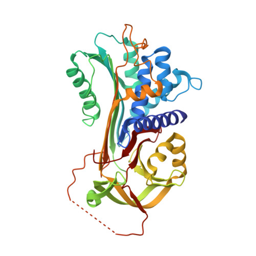
The hormones that most directly control tissue activities in health and disease are delivered by two noninhibitory members of the serpin family of protease inhibitors, thyroxine-binding globulin (TBG) and corticosteroid-binding globulin. The structure of TBG bound to tetra-iodo thyroxine, solved here at 2.8 A, shows how the thyroxine is carried in a surface pocket on the molecule. This unexpected binding site is confirmed by mutations associated with a loss of hormone binding in both TBG and also homologously in corticosteroid-binding globulin. TBG strikingly differs from other serpins in having the upper half of its main beta-sheet fully opened, so its reactive center peptide loop can readily move in and out of the sheet to give an equilibrated binding and release of thyroxine. The entry of the loop triggers a conformational change, with a linked contraction of the binding pocket and release of the bound thyroxine. The ready reversibility of this change is due to the unique presence in the reactive loop of TBG of a proline that impedes the full and irreversible entry of the loop that occurs in other serpins. Thus, TBG has adapted the serpin inhibitory mechanism to give a reversible flip-flop transition, from a high-affinity to a low-affinity form. The complexity and ready triggering of this conformational mechanism strongly indicates that TBG has evolved to allow a modulated and targeted delivery of thyroxine to the tissues.
Organizational Affiliation:
Departments of Haematology and Medicine, Cambridge Institute for Medical Research, University of Cambridge, Hills Road, Cambridge CB2 2XY, United Kingdom.