The Crystal Structure and Mutational Binding Analysis of the Extracellular Domain of the Platelet-Activating Receptor Clec-2.
Watson, A.A., Brown, J., Harlos, K., Eble, J.A., Walter, T.S., O'Callaghan, C.A.(2007) J Biol Chem 282: 3165
- PubMed: 17132623
- DOI: https://doi.org/10.1074/jbc.M610383200
- Primary Citation of Related Structures:
2C6U - PubMed Abstract:
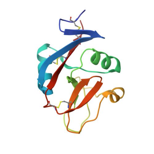
The human C-type lectin-like molecule CLEC-2 is expressed on the surface of platelets and signaling through CLEC-2 causes platelet activation and aggregation. CLEC-2 is a receptor for the platelet-aggregating snake venom protein rhodocytin. It is also a newly identified co-receptor for human immunodeficiency virus type 1 (HIV-1). An endogenous ligand has not yet been identified. We have solved the crystal structure of the extracellular domain of CLEC-2 to 1.6-A resolution, and identified the key structural features involved in ligand binding. A semi-helical loop region and flanking residues dominate the surface that is available for ligand binding. The precise distribution of hydrophobic and electrostatic features in this loop will determine the nature of any endogenous ligand with which it can interact. Major ligand-induced conformational change in CLEC-2 is unlikely as its overall fold is compact and robust. However, ligand binding could induce a tilt of a 3-10 helical portion of the long loop region. Mutational analysis and surface plasmon resonance binding studies support these observations. This study provides a framework for understanding the effects of rhodocytin venom binding on CLEC-2 and for understanding the nature of likely endogenous ligands and will provide a basis for rational design of drugs to block ligand binding.
Organizational Affiliation:
Henry Wellcome Building for Molecular Physiology, Division of Structural Biology, University of Oxford, Roosevelt Drive, Oxford OX3 7BN, United Kingdom.